 Explore
 Validate
 Learn
Learn Western blot
Antibody data
- Antibody Data
- Antigen structure
- References [7]
- Comments [0]
- Validations
- Western blot [3]
- Immunocytochemistry [7]
- Immunoprecipitation [1]
- Immunohistochemistry [3]
- Chromatin Immunoprecipitation [2]
- Other assay [1]
Submit
Validation data
Reference
Comment
Report error
- Product number
- MA1-028 - Provider product page

- Provider
- Invitrogen Antibodies
- Product name
- GATA3 Monoclonal Antibody (1A12-1D9)
- Antibody type
- Monoclonal
- Antigen
- Purifed from natural sources
- Description
- Western blot analysis of MA1-028 detects a prominent ~50 kDa protein in GATA3 expressing SH-SY5Y cells. The signal is enhanced in nuclear extracts due to enrichment of GATA3. This antibody has also been successfully tested in IP, IHC and Immunofluorescence applications. MA1-028 can be used for immunofluoresence analysis of Gata3 in human ESC lines. MA1-028 was successfully used in western blotting analysis of GATA3 in breast cancer cell lines.
- Reactivity
- Human, Mouse, Rat
- Host
- Mouse
- Isotype
- IgG
- Antibody clone number
- 1A12-1D9
- Vial size
- 100 μg
- Concentration
- 1 mg/mL
- Storage
- -20°C
Submitted references Krüppel-like factor 5 rewires NANOG regulatory network to activate human naive pluripotency specific LTR7Ys and promote naive pluripotency.
Naive stem cell blastocyst model captures human embryo lineage segregation.
Assessment of aneuploidy concordance between clinical trophectoderm biopsy and blastocyst.
Highly multiplexed immunofluorescence images and single-cell data of immune markers in tonsil and lung cancer.
Selective gene dependencies in MYCN-amplified neuroblastoma include the core transcriptional regulatory circuitry.
Improving Cell Survival in Injected Embryos Allows Primed Pluripotent Stem Cells to Generate Chimeric Cynomolgus Monkeys.
Self-organization of the in vitro attached human embryo.
Ai Z, Xiang X, Xiang Y, Szczerbinska I, Qian Y, Xu X, Ma C, Su Y, Gao B, Shen H, Bin Ramli MN, Chen D, Liu Y, Hao JJ, Ng HH, Zhang D, Chan YS, Liu W, Liang H
Cell reports 2022 Aug 23;40(8):111240
Cell reports 2022 Aug 23;40(8):111240
Naive stem cell blastocyst model captures human embryo lineage segregation.
Yanagida A, Spindlow D, Nichols J, Dattani A, Smith A, Guo G
Cell stem cell 2021 Jun 3;28(6):1016-1022.e4
Cell stem cell 2021 Jun 3;28(6):1016-1022.e4
Assessment of aneuploidy concordance between clinical trophectoderm biopsy and blastocyst.
Victor AR, Griffin DK, Brake AJ, Tyndall JC, Murphy AE, Lepkowsky LT, Lal A, Zouves CG, Barnes FL, McCoy RC, Viotti M
Human reproduction (Oxford, England) 2019 Jan 1;34(1):181-192
Human reproduction (Oxford, England) 2019 Jan 1;34(1):181-192
Highly multiplexed immunofluorescence images and single-cell data of immune markers in tonsil and lung cancer.
Rashid R, Gaglia G, Chen YA, Lin JR, Du Z, Maliga Z, Schapiro D, Yapp C, Muhlich J, Sokolov A, Sorger P, Santagata S
Scientific data 2019 Dec 17;6(1):323
Scientific data 2019 Dec 17;6(1):323
Selective gene dependencies in MYCN-amplified neuroblastoma include the core transcriptional regulatory circuitry.
Durbin AD, Zimmerman MW, Dharia NV, Abraham BJ, Iniguez AB, Weichert-Leahey N, He S, Krill-Burger JM, Root DE, Vazquez F, Tsherniak A, Hahn WC, Golub TR, Young RA, Look AT, Stegmaier K
Nature genetics 2018 Sep;50(9):1240-1246
Nature genetics 2018 Sep;50(9):1240-1246
Improving Cell Survival in Injected Embryos Allows Primed Pluripotent Stem Cells to Generate Chimeric Cynomolgus Monkeys.
Kang Y, Ai Z, Duan K, Si C, Wang Y, Zheng Y, He J, Yin Y, Zhao S, Niu B, Zhu X, Liu L, Xiang L, Zhang L, Niu Y, Ji W, Li T
Cell reports 2018 Nov 27;25(9):2563-2576.e9
Cell reports 2018 Nov 27;25(9):2563-2576.e9
Self-organization of the in vitro attached human embryo.
Deglincerti A, Croft GF, Pietila LN, Zernicka-Goetz M, Siggia ED, Brivanlou AH
Nature 2016 May 12;533(7602):251-4
Nature 2016 May 12;533(7602):251-4
No comments: Submit comment
Supportive validation
- Submitted by
- Invitrogen Antibodies (provider)
- Main image

- Experimental details
- Western blot analysis of GATA3 was performed by loading 15 µg of HEK293T cell lysate overexpressing GATA3-DDK (right lane) or empty vector control (left lane), and 10 µL PageRuler Plus Prestained Protein Ladder (Product # 26619) onto a 4-20% Tris-HCl polyacrylamide gel. Proteins were transferred to a PVDF membrane and blocked with StartingBlock T20 (Product # 37543) for at least 1 hour. The membrane was probed with a GATA3 monoclonal antibody (Product # MA1-028) at a dilution of 1:1000 overnight at 4°C on a rocking platform, washed in TBS-0.1%Tween-20, and probed with an HRP-conjugated goat anti-mouse IgG secondary antibody (Product # 31430) at a dilution of 1:20,000 for 1 hour. Chemiluminescent detection was performed using SuperSignal West Dura (Product # 34075). Images were acquired on a Thermo Scientific myECL Imager (Product # 62236).

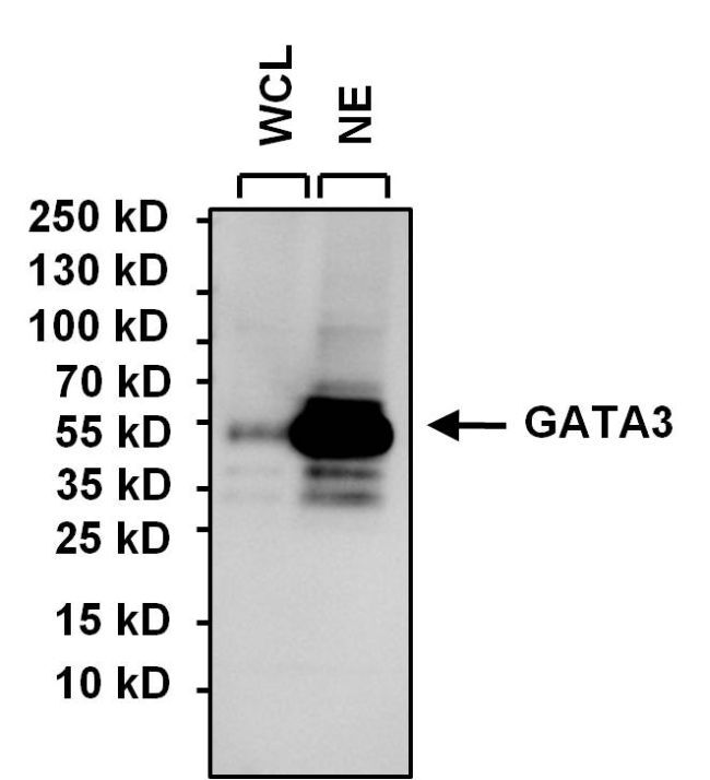
- Submitted by
- Invitrogen Antibodies (provider)
- Main image

- Experimental details
- Western blot analysis of GATA3 was performed by loading 15 µg of SH-SY5Y whole cell lysate (WCL), SH-SY5Y nuclear extract (NE) and 10 µL of PageRuler Plus Prestained Protein Ladder (Product # 26619) onto a 4-20% Tris-HCl polyacrylamide gel. Proteins were transferred to a PVDF membrane and blocked with StartingBlock T20 (Product # 37543) for at least 1 hour. The membrane was probed with a GATA3 monoclonal antibody (Product # MA1-028) at a dilution of 1:1000 overnight at 4°C on a rocking platform, washed in TBS-0.1%Tween-20, and probed with an HRP-conjugated goat anti-mouse IgG secondary antibody (Product # 31430) at a dilution of 1:20,000 for 1 hour. Chemiluminescent detection was performed using SuperSignal West Dura (Product # 34075). Images were acquired on a Thermo Scientific myECL Imager (Product # 62236). Note: Nuclear extracts were obtained using the Thermo Scientific NE-PER Nuclear and Cytoplasmic Extraction Kit (Product # 78833).
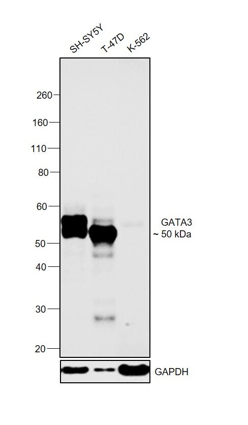
- Submitted by
- Invitrogen Antibodies (provider)
- Main image
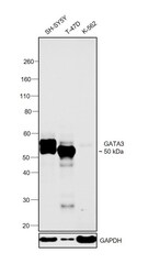
- Experimental details
- Western blot was performed using Anti-GATA3 Monoclonal Antibody (1A12-1D9) (Product # MA1-028) and a 50 kDa band corresponding to GATA3 was observed across cell lines tested except K-562 which is reported to be negative. Modified whole cell extracts (1% SDS) (30 µg lysate) of SH-SY5Y (Lane 1), T-47D (Lane 2) and K-562 (Lane 3) were electrophoresed using NuPAGE™ 4-12% Bis-Tris Protein Gel (Product # NP0322BOX). Resolved proteins were then transferred onto a nitrocellulose membrane (Product # IB23001) by iBlot® 2 Dry Blotting System (Product # IB21001). The blot was probed with the primary antibody (1:1000 dilution) and detected by chemiluminescence with Goat anti-Mouse IgG (H+L) Superclonal™ Recombinant Secondary Antibody, HRP (Product # A28177, 1:4000 dilution) using the iBright FL 1000 (Product # A32752). Chemiluminescent detection was performed using SuperSignal™ West Dura Extended Duration Substrate (Product # 34076).
Supportive validation
- Submitted by
- Invitrogen Antibodies (provider)
- Main image
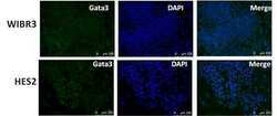
- Experimental details
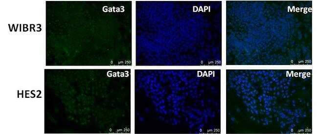
- Immunofluorescent analysis of Gata3 (green) in human ESCs WIRB3 and HES2. The cells were fixed with 300 uL 4% paraformaldehyde solution for 15 min at RT in the dark, permeabilized with 300 uL 0.25% TritonX-100 solution (diluted in PBS) for 5 min at RT, and blocked with 300 uL 10% BSA solution (diluted in PBS) for 30 min in a 37C incubator. Cells were stained with a Gata3 monoclonal antibody (Product # MA1-028) at a dilution of 1:100 (2 µg/mL) in 3% BSA solution (diluted in PBS) at 4C overnight, and then incubated with a secondary antibody (donkey anti mouse-AlexaFluor 488, green). Nuclei (blue) was stained with DAPI (Product # D1306). Images were taken on a Leica DM5500 microscope at 10X magnification. Data courtesy of Dr. Jianlong Wang's lab.
- Submitted by
- Invitrogen Antibodies (provider)
- Main image
- Experimental details
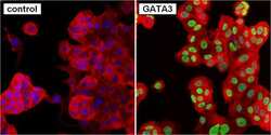
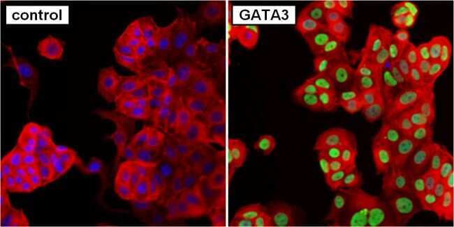
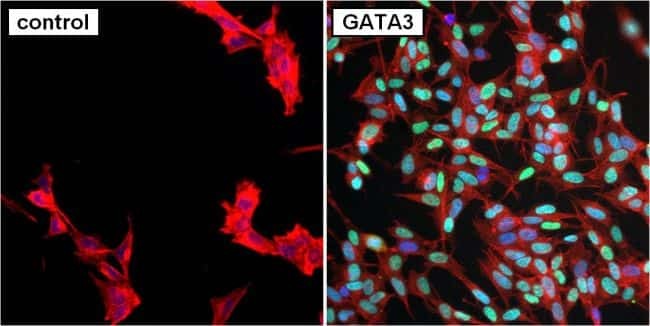
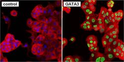
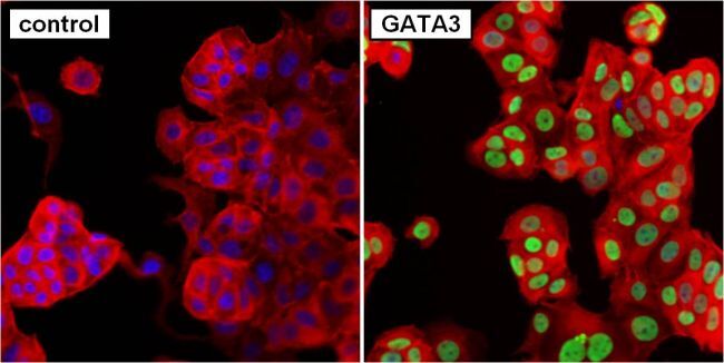
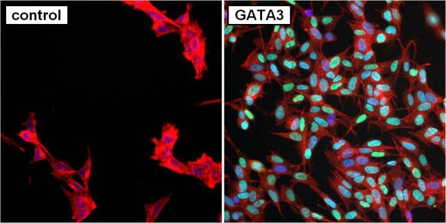
- Immunofluorescent analysis of GATA3 (green) in MCF7 cells. Formalin fixed cells were permeabilized with 0.1% Triton X-100 in TBS for 10 minutes at room temperature. Cells were blocked with 1% Blocker BSA (Product # 37525) for 15 minutes at room temperature. Cells were probed without (left panel) or with (right panel) a GATA3 monoclonal antibody (Product # MA1-028) at a dilution of 1:50 for at least 1 hour at room temperature, washed with PBS, and incubated with a DyLight 488-conjugated goat anti-mouse IgG secondary antibody (Product # 35503) at a dilution of 1:400 for 30 minutes at room temperature. F-Actin (red) was stained with DyLight-554 Phalloidin (Product # 21834) and nuclei (blue) were stained with Hoechst 33342 dye (Product # 62249). Images were taken on a Thermo Scientific ToxInsight Instrument at 20X magnification.
- Submitted by
- Invitrogen Antibodies (provider)
- Main image

- Experimental details
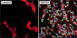
- Immunofluorescent analysis of GATA3 (green) in SH-SY5Y cells. Formalin fixed cells were permeabilized with 0.1% Triton X-100 in TBS for 10 minutes at room temperature. Cells were blocked with 1% Blocker BSA (Product # 37525) for 15 minutes at room temperature. Cells were probed without (left panel) or with (right panel) a GATA3 monoclonal antibody (Product # MA1-028) at a dilution of 1:50 for at least 1 hour at room temperature, washed with PBS, and incubated with a DyLight 488-conjugated goat anti-mouse IgG secondary antibody (Product # 35503) at a dilution of 1:400 for 30 minutes at room temperature. F-Actin (red) was stained with DyLight-554 Phalloidin (Product # 21834) and nuclei (blue) were stained with Hoechst 33342 dye (Product # 62249). Images were taken on a Thermo Scientific ToxInsight Instrument at 20X magnification.
- Submitted by
- Invitrogen Antibodies (provider)
- Main image

- Experimental details
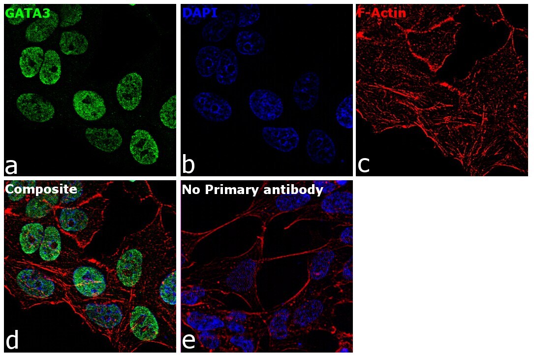
- Immunofluorescence analysis of GATA3 was performed using 70% confluent log phase MCF7 cells. The cells were fixed with 4% paraformaldehyde for 10 minutes, permeabilized with 0.1% Triton™ X-100 for 15 minutes, and blocked with 2% BSA for 1 hour at room temperature. The cells were labeled with GATA3 Mouse Monoclonal Antibody (1A12-1D9) (Product # MA1-028) at 5 µg/mL in 0.1% BSA, incubated at 4 degree celsius overnight and then with Goat anti-Mouse IgG (H+L) Superclonal™ Recombinant Secondary Antibody, Alexa Fluor® 488 conjugate (Product # A28175) at a dilution of 1:2000 for 45 minutes at room temperature (Panel a: green). Nuclei (Panel b: blue) were stained with ProLong™ Diamond Antifade Mountant with DAPI (Product # P36962). F-actin (Panel c: red) was stained with Rhodamine Phalloidin (Product # R415, 1:300). Panel d represents the merged image showing Nuclear localization. Panel e represents control cells with no primary antibody to assess background. The images were captured at 60X magnification.
- Submitted by
- Invitrogen Antibodies (provider)
- Main image

- Experimental details
- Immunofluorescence analysis of GATA3 was performed using 70% confluent log phase MCF7 cells. The cells were fixed with 4% paraformaldehyde for 10 minutes, permeabilized with 0.1% Triton™ X-100 for 15 minutes, and blocked with 2% BSA for 1 hour at room temperature. The cells were labeled with GATA3 Mouse Monoclonal Antibody (1A12-1D9) (Product # MA1-028) at 5 µg/mL in 0.1% BSA, incubated at 4 degree celsius overnight and then with Goat anti-Mouse IgG (H+L) Superclonal™ Recombinant Secondary Antibody, Alexa Fluor® 488 conjugate (Product # A28175) at a dilution of 1:2000 for 45 minutes at room temperature (Panel a: green). Nuclei (Panel b: blue) were stained with ProLong™ Diamond Antifade Mountant with DAPI (Product # P36962). F-actin (Panel c: red) was stained with Rhodamine Phalloidin (Product # R415, 1:300). Panel d represents the merged image showing Nuclear localization. Panel e represents control cells with no primary antibody to assess background. The images were captured at 60X magnification.

- Submitted by
- Invitrogen Antibodies (provider)
- Main image
- Experimental details
- Immunofluorescent analysis of GATA3 (green) in MCF7 cells. Formalin fixed cells were permeabilized with 0.1% Triton X-100 in TBS for 10 minutes at room temperature. Cells were blocked with 1% Blocker BSA (Product # 37525) for 15 minutes at room temperature. Cells were probed without (left panel) or with (right panel) a GATA3 monoclonal antibody (Product # MA1-028) at a dilution of 1:50 for at least 1 hour at room temperature, washed with PBS, and incubated with a DyLight 488-conjugated goat anti-mouse IgG secondary antibody (Product # 35503) at a dilution of 1:400 for 30 minutes at room temperature. F-Actin (red) was stained with DyLight-554 Phalloidin (Product # 21834) and nuclei (blue) were stained with Hoechst 33342 dye (Product # 62249). Images were taken on a Thermo Scientific ToxInsight Instrument at 20X magnification.
- Submitted by
- Invitrogen Antibodies (provider)
- Main image
- Experimental details
- Immunofluorescent analysis of GATA3 (green) in SH-SY5Y cells. Formalin fixed cells were permeabilized with 0.1% Triton X-100 in TBS for 10 minutes at room temperature. Cells were blocked with 1% Blocker BSA (Product # 37525) for 15 minutes at room temperature. Cells were probed without (left panel) or with (right panel) a GATA3 monoclonal antibody (Product # MA1-028) at a dilution of 1:50 for at least 1 hour at room temperature, washed with PBS, and incubated with a DyLight 488-conjugated goat anti-mouse IgG secondary antibody (Product # 35503) at a dilution of 1:400 for 30 minutes at room temperature. F-Actin (red) was stained with DyLight-554 Phalloidin (Product # 21834) and nuclei (blue) were stained with Hoechst 33342 dye (Product # 62249). Images were taken on a Thermo Scientific ToxInsight Instrument at 20X magnification.
Supportive validation
- Submitted by
- Invitrogen Antibodies (provider)
- Main image

- Experimental details
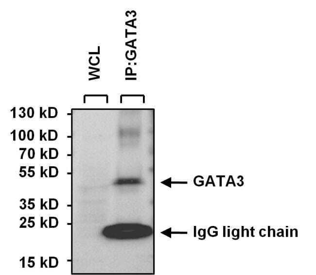
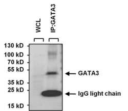
- Immunoprecipitation of GATA3 was performed on SH-SY5Y cells. Antigen-antibody complexes were formed by incubating 250 µg of SH-SY5Y whole cell lysate with 5 µg of a GATA3 monoclonal antibody (Product # MA1-028) overnight on a rocking platform at 4°C. The immune complexes were captured on 50 µL Protein A/G Agarose (Product # 20421), washed extensively, and eluted with 5X Lane Marker Reducing Sample Buffer (Product # 39000). Eluted sample and 25 µg of SH-SY5Y whole cell lysate (loading control) were resolved on a 4-20% Tris-HCl polyacrylamide gel, transferred to a PVDF membrane, and blocked with StartingBlock T20 (Product # 37543) for at least 1 hour. The membrane was probed with a GATA3 monoclonal antibody (Product # MA1-028) at a dilution of 1:1000 overnight rotating at 4°C, washed in TBST, and probed with an HRP- conjugated, light chain specific goat anti-mouse IgG at a dilution of 1:20,000 for at least 1 hour. Chemiluminescent detection was performed using SuperSignal West Pico (Product # 34080). Images were acquired on a Thermo Scientific myECL Imager (Product # 62236).
Supportive validation
- Submitted by
- Invitrogen Antibodies (provider)
- Main image
- Experimental details
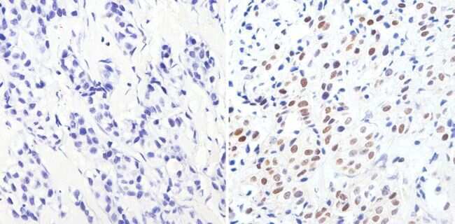
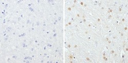
- Immunohistochemistry analysis of GATA3 showing staining in the nucleus of paraffin-embedded human breast carcinoma (right) compared to a negative control without primary antibody (left). To expose target proteins, antigen retrieval was performed using 10mM sodium citrate (pH 6.0), microwaved for 8-15 min. Following antigen retrieval, tissues were blocked in 3% H2O2-methanol for 15 min at room temperature, washed with ddH2O and PBS, and then probed with a GATA3 monoclonal antibody (Product # MA1-028) diluted in 3% BSA-PBS at a dilution of 1:1000 overnight at 4°C in a humidified chamber. Tissues were washed extensively in PBST and detection was performed using an HRP-conjugated secondary antibody followed by colorimetric detection using a DAB kit. Tissues were counterstained with hematoxylin and dehydrated with ethanol and xylene to prep for mounting.
- Submitted by
- Invitrogen Antibodies (provider)
- Main image
- Experimental details
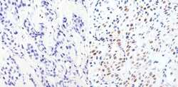
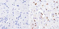
- Immunohistochemistry analysis of GATA3 showing staining in the nucleus of paraffin-embedded mouse brain tissue (right) compared to a negative control without primary antibody (left). To expose target proteins, antigen retrieval was performed using 10mM sodium citrate (pH 6.0), microwaved for 8-15 min. Following antigen retrieval, tissues were blocked in 3% H2O2-methanol for 15 min at room temperature, washed with ddH2O and PBS, and then probed with a GATA3 monoclonal antibody (Product # MA1-028) diluted in 3% BSA-PBS at a dilution of 1:1000 overnight at 4°C in a humidified chamber. Tissues were washed extensively in PBST and detection was performed using an HRP-conjugated secondary antibody followed by colorimetric detection using a DAB kit. Tissues were counterstained with hematoxylin and dehydrated with ethanol and xylene to prep for mounting.

- Submitted by
- Invitrogen Antibodies (provider)
- Main image
- Experimental details
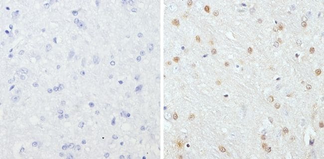
- Immunohistochemistry analysis of GATA3 showing staining in the nucleus of paraffin-embedded rat brain tissue (right) compared to a negative control without primary antibody (left). To expose target proteins, antigen retrieval was performed using 10mM sodium citrate (pH 6.0), microwaved for 8-15 min. Following antigen retrieval, tissues were blocked in 3% H2O2-methanol for 15 min at room temperature, washed with ddH2O and PBS, and then probed with a GATA3 monoclonal antibody (Product # MA1-028) diluted in 3% BSA-PBS at a dilution of 1:200 overnight at 4°C in a humidified chamber. Tissues were washed extensively in PBST and detection was performed using an HRP-conjugated secondary antibody followed by colorimetric detection using a DAB kit. Tissues were counterstained with hematoxylin and dehydrated with ethanol and xylene to prep for mounting.
Supportive validation
- Submitted by
- Invitrogen Antibodies (provider)
- Main image
- Experimental details
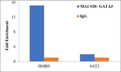
- Chromatin Immunoprecipitation (ChIP) assay of endogenous GATA3 protein using Anti-GATA3 Antibody: ChIP was performed using Anti-GATA3 Monoclonal Antibody (1A12-1D9) (Product # MA1-028) 5 µg, on sheared chromatin from HCT116 cells using the MAGnify ChIP System kit (Product # 49-2024). Normal Mouse IgG was used as a negative IP control. The purified DNA was analyzed by qPCR using primers binding to promoter of HMBS and SAT2 satellite repeats. Data is presented as fold enrichment of the antibody signal versus the negative control IgG using the comparative CT method.

- Submitted by
- Invitrogen Antibodies (provider)
- Main image

- Experimental details
- Chromatin Immunoprecipitation (ChIP) assay of endogenous GATA3 protein using Anti-GATA3 Antibody: ChIP was performed using Anti-GATA3 Monoclonal Antibody (1A12-1D9) (Product # MA1-028) 5 µg, on sheared chromatin from HCT116 cells using the MAGnify ChIP System kit (Product # 49-2024). Normal Mouse IgG was used as a negative IP control. The purified DNA was analyzed by qPCR using primers binding to promoter of HMBS and SAT2 satellite repeats. Data is presented as fold enrichment of the antibody signal versus the negative control IgG using the comparative CT method.

Supportive validation
- Submitted by
- Invitrogen Antibodies (provider)
- Main image
- Experimental details
- Immunoprecipitation of GATA3 was performed on SH-SY5Y cells. Antigen-antibody complexes were formed by incubating 250 µg of SH-SY5Y whole cell lysate with 5 µg of a GATA3 monoclonal antibody (Product # MA1-028) overnight on a rocking platform at 4øC. The immune complexes were captured on 50 µL Protein A/G Agarose (Product # 20421), washed extensively, and eluted with 5X Lane Marker Reducing Sample Buffer (Product # 39000). Eluted sample and 25 µg of SH-SY5Y whole cell lysate (loading control) were resolved on a 4-20% Tris-HCl polyacrylamide gel, transferred to a PVDF membrane, and blocked with StartingBlock T20 (Product # 37543) for at least 1 hour. The membrane was probed with a GATA3 monoclonal antibody (Product # MA1-028) at a dilution of 1:1000 overnight rotating at 4øC, washed in TBST, and probed with an HRP- conjugated, light chain specific goat anti-mouse IgG at a dilution of 1:20,000 for at least 1 hour. Chemiluminescent detection was performed using SuperSignal West Pico (Product # 34080). Images were acquired on a Thermo Scientific myECL Imager (Product # 62236).
