Explore
 Validate
 Learn
Learn Western blot
Antibody data
- Antibody Data
- Antigen structure
- References [2]
- Comments [0]
- Validations
- Western blot [2]
- Immunocytochemistry [2]
- Immunoprecipitation [1]
- Immunohistochemistry [3]
Submit
Validation data
Reference
Comment
Report error
- Product number
- GTX30600 - Provider product page

- Provider
- GeneTex
- Product name
- GATA3 antibody [1A12-1D9]
- Antibody type
- Monoclonal
- Reactivity
- Human, Mouse, Rat
- Host
- Mouse
Submitted references The impact of the effectiveness of GATA3 as a prognostic factor in breast cancer.
The FOXN3-NEAT1-SIN3A repressor complex promotes progression of hormonally responsive breast cancer.
Fararjeh AS, Tu SH, Chen LC, Liu YR, Lin YK, Chang HL, Chang HW, Wu CH, Hwang-Verslues WW, Ho YS
Human pathology 2018 Oct;80:219-230
Human pathology 2018 Oct;80:219-230
The FOXN3-NEAT1-SIN3A repressor complex promotes progression of hormonally responsive breast cancer.
Li W, Zhang Z, Liu X, Cheng X, Zhang Y, Han X, Zhang Y, Liu S, Yang J, Xu B, He L, Sun L, Liang J, Shang Y
The Journal of clinical investigation 2017 Sep 1;127(9):3421-3440
The Journal of clinical investigation 2017 Sep 1;127(9):3421-3440
No comments: Submit comment
Supportive validation
- Submitted by
- GeneTex (provider)
- Main image

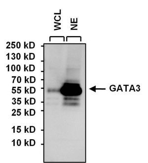
- Experimental details
- WB analysis of SH-SY5Y whole cell lysate (WCL) and SH-SY5Y nuclear extract (NE) (15ug per lane) using GATA3 antibody [1A12-1D9] at a dilution of 1:1000.
- Submitted by
- GeneTex (provider)
- Main image
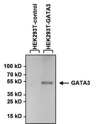
- Experimental details
- WB analysis of lysates from HEK293T cells overexpressing GATA3-DDK (right) or empty vector control (left) using GATA3 antibody [1A12-1D9] at a dilution of 1:1000.

Supportive validation
- Submitted by
- GeneTex (provider)
- Main image
- Experimental details
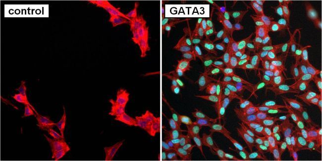
- ICC/IF analysis of SH-SY5Y cells without (left) or with (right) GATA3 antibody [1A12-1D9] at a dilution of 1:50 (green). F-Actin (red) was stained with Phalloidin and nuclei (blue) were stained with Hoechst 33342.
- Submitted by
- GeneTex (provider)
- Main image
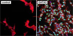
- Experimental details
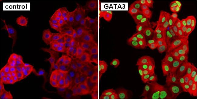
- ICC/IF analysis of MCF7 cells without (left) or with (right) GATA3 antibody [1A12-1D9] at a dilution of 1:50 (green). F-Actin (red) was stained with Phalloidin and nuclei (blue) were stained with Hoechst 33342.
Supportive validation
- Submitted by
- GeneTex (provider)
- Main image
- Experimental details
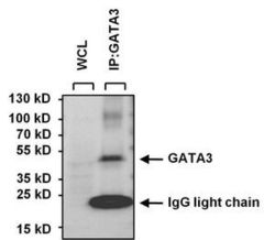
- IP analysis was performed with 250ug of SH-SY5Y whole cell lysate and 5ug of GATA3 antibody [1A12-1D9]. The precipitate and 25ug of SH-SY5Y whole cell lysate (loading control) were detected by the same antibody at a dilution of 1:1000.

Supportive validation
- Submitted by
- GeneTex (provider)
- Main image
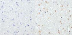
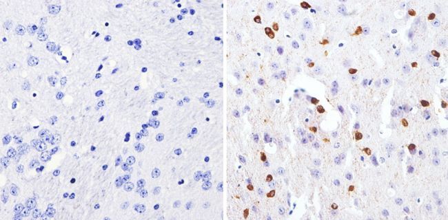
- Experimental details
- IHC-P analysis of rat brain tissues with (right) or without GATA3 antibody [1A12-1D9] at a dilution of 1:200. To expose target proteins, antigen retrieval was performed using 10mM sodium citrate (pH 6.0), microwaved for 8-15 min.
- Submitted by
- GeneTex (provider)
- Main image
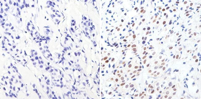
- Experimental details
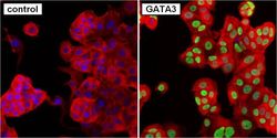
- IHC-P analysis of human breast carcinoma with (right) or without (left) GATA3 antibody [1A12-1D9] at a dilution of 1:1000. To expose target proteins, antigen retrieval was performed using 10mM sodium citrate (pH 6.0), microwaved for 8-15 min.
- Submitted by
- GeneTex (provider)
- Main image

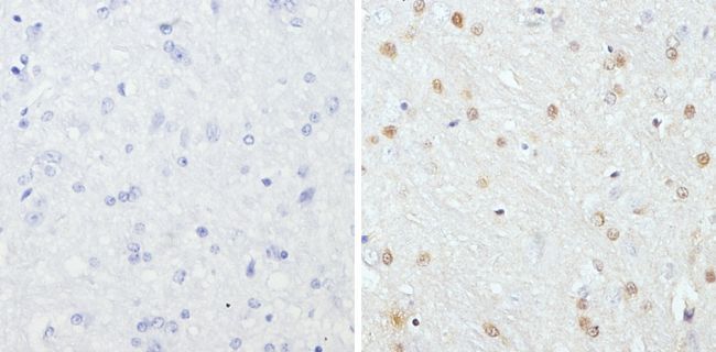
- Experimental details
- IHC-P analysis of mouse brain tissues with (right) or without (left) GATA3 antibody [1A12-1D9] at a dilution of 1:1000. To expose target proteins, antigen retrieval was performed using 10mM sodium citrate (pH 6.0), microwaved for 8-15 min.