Explore
 Validate
 Learn
Learn Western blot
Antibody data
- Antibody Data
- Antigen structure
- References [4]
- Comments [0]
- Validations
- Western blot [2]
- Immunocytochemistry [1]
- Immunohistochemistry [1]
- Other assay [1]
Submit
Validation data
Reference
Comment
Report error
- Product number
- MAB6330 - Provider product page

- Provider
- R&D Systems
- Product name
- Human/Mouse GATA-3 Antibody
- Antibody type
- Monoclonal
- Description
- Protein A or G purified from hybridoma culture supernatant. Detects human and mouse GATA-3 in Western blots.
- Reactivity
- Human, Mouse
- Host
- Mouse
- Conjugate
- Unconjugated
- Antigen sequence
P23771- Isotype
- IgG
- Antibody clone number
- 634913
- Vial size
- 100 ug
- Concentration
- LYOPH
- Storage
- Use a manual defrost freezer and avoid repeated freeze-thaw cycles. 12 months from date of receipt, -20 to -70 °C as supplied. 1 month, 2 to 8 °C under sterile conditions after reconstitution. 6 months, -20 to -70 °C under sterile conditions after reconstitution.
Submitted references Increased TREM-2 expression on the subsets of CD11c+ cells in the lungs and lymph nodes during allergic airway inflammation.
ZNF503/Zpo2 drives aggressive breast cancer progression by down-regulation of GATA3 expression.
Analysis of histological and immunological parameters of metastatic lymph nodes from colon cancer patients reveals that T-helper 1 type immune response is associated with improved overall survival.
Methylation of Gata3 protein at Arg-261 regulates transactivation of the Il5 gene in T helper 2 cells.
Hall SC, Agrawal DK
Scientific reports 2017 Sep 19;7(1):11853
Scientific reports 2017 Sep 19;7(1):11853
ZNF503/Zpo2 drives aggressive breast cancer progression by down-regulation of GATA3 expression.
Shahi P, Wang CY, Lawson DA, Slorach EM, Lu A, Yu Y, Lai MD, Gonzalez Velozo H, Werb Z
Proceedings of the National Academy of Sciences of the United States of America 2017 Mar 21;114(12):3169-3174
Proceedings of the National Academy of Sciences of the United States of America 2017 Mar 21;114(12):3169-3174
Analysis of histological and immunological parameters of metastatic lymph nodes from colon cancer patients reveals that T-helper 1 type immune response is associated with improved overall survival.
Nizri E, Greenman-Maaravi N, Bar-David S, Ben-Yehuda A, Weiner G, Lahat G, Klausner J
Medicine 2016 Nov;95(45):e5340
Medicine 2016 Nov;95(45):e5340
Methylation of Gata3 protein at Arg-261 regulates transactivation of the Il5 gene in T helper 2 cells.
Hosokawa H, Kato M, Tohyama H, Tamaki Y, Endo Y, Kimura MY, Tumes DJ, Motohashi S, Matsumoto M, Nakayama KI, Tanaka T, Nakayama T
The Journal of biological chemistry 2015 May 22;290(21):13095-103
The Journal of biological chemistry 2015 May 22;290(21):13095-103
No comments: Submit comment
Supportive validation
- Submitted by
- R&D Systems (provider)
- Main image
- Experimental details
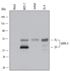
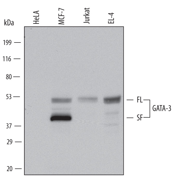
- Detection of Human and Mouse GATA-3 by Western Blot. Western blot shows lysates of HeLa human cervical epithelial carcinoma cell line, MCF-7 human breast cancer cell line, Jurkat human acute T cell leukemia cell line, and EL-4 mouse lymphoblast cell line. PVDF Membrane was probed with 0.1 µg/mL of Moue Anti-Human/Mouse GATA-3 Monoclonal Antibody (Catalog # MAB6330) followed by HRP-conjugated Anti-Mouse IgG Secondary Antibody (Catalog # HAF007). Specific bands were detected for full length (FL) GATA-3 at approximately 52 kDa and the splice form (SF) found in MCF-7 cells at approximately 40 kDa (as indicated). This experiment was conducted under reducing conditions and using Immunoblot Buffer Group 1.
- Submitted by
- R&D Systems (provider)
- Main image
- Experimental details
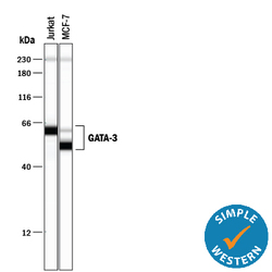
- Detection of Human GATA-3 by Simple WesternTM. Simple Western lane view shows lysates of Jurkat human acute T cell leukemia cell line and MCF-7 human breast cancer cell line, loaded at 0.5 mg/mL. Specific bands were detected for GATA-3 at approximately 53 (splice variant) and 61 kDa (full length) as indicated, using 10 µg/mL of Mouse Anti-Human/Mouse GATA-3 Monoclonal Antibody (Catalog # MAB6330). This experiment was conducted under reducing conditions and using the 12-230 kDa separation system. Non-specific interaction with the 230 kDa Simple Western standard may be seen with this antibody.

Supportive validation
- Submitted by
- R&D Systems (provider)
- Main image
- Experimental details
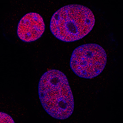
- GATA-3 in MCF-7 Human Cell Line. GATA-3 was detected in immersion fixed MCF-7 human breast cancer cell line using Mouse Anti-Human/Mouse GATA-3 Monoclonal Antibody (Catalog # MAB6330) at 3 µg/mL for 3 hours at room temperature. Cells were stained using the NorthernLights™ 557-conjugated Anti-Mouse IgG Secondary Antibody (red; Catalog # NL007) and counterstained with DAPI (blue). Specific staining was localized to nuclei. View our protocol for Fluorescent ICC Staining of Cells on Coverslips.

Supportive validation
- Submitted by
- R&D Systems (provider)
- Main image
- Experimental details
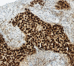
- GATA-3 in Human Breast Cancer Tissue. GATA-3 was detected in immersion fixed paraffin-embedded sections of human breast cancer tissue using Mouse Anti-Human/Mouse GATA-3 Monoclonal Antibody (Catalog # MAB6330) at 15 µg/mL overnight at 4 °C. Before incubation with the primary antibody, tissue was subjected to heat-induced epitope retrieval using Antigen Retrieval Reagent-Basic (Catalog # CTS013). Tissue was stained using the Anti-Mouse HRP-DAB Cell & Tissue Staining Kit (brown; Catalog # CTS002) and counterstained with hematoxylin (blue). Specific staining was localized to nuclei. View our protocol for Chromogenic IHC Staining of Paraffin-embedded Tissue Sections.

Supportive validation
- Submitted by
- R&D Systems (provider)
- Main image

- Experimental details
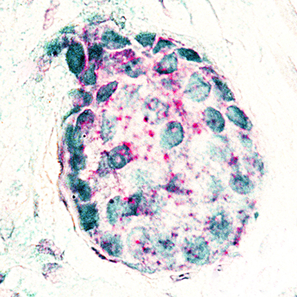
- GATA-3 in Human Breast Cancer Tissue Using Dual RNAscope® ISH and IHC. GATA-3 mRNA (red) and protein (green) was detected in formalin-fixed paraffin-embedded tissue sections of human breast cancer tissue probed with ACD RNAScope® Probe (Catalog # 403551) followed by immunohistochemistry using R&D Systems Mouse Anti-Human/Mouse GATA-3 Monoclonal Antibody (Catalog# MAB6330) at 5ug/mL for 1 hour at room temperature followed by incubation with the Anti-Mouse IgG VisUCyte HRP Polymer Antibody (R&D Systems, Catalog # VC001). Tissue was stained using ACD RNAscope® 2.5 HD Duplex Detection Reagents (Catalog # 322500).