Explore
 Validate
 Learn
LearnMA5-25345
antibody from Invitrogen Antibodies
Targeting: LMAN1
ERGIC-53, ERGIC53, F5F8D, FMFD1, gp58, MCFD1, MR60
 Western blot
 Immunocytochemistry
 Flow cytometry
Antibody data
- Antibody Data
- Antigen structure
- References [1]
- Comments [0]
- Validations
- Immunocytochemistry [3]
- Immunohistochemistry [6]
- Other assay [1]
Submit
Validation data
Reference
Comment
Report error
- Product number
- MA5-25345 - Provider product page

- Provider
- Invitrogen Antibodies
- Product name
- LMAN1 Monoclonal Antibody (OTI1A8)
- Antibody type
- Monoclonal
- Antigen
- Recombinant full-length protein
- Reactivity
- Human, Rat, Canine
- Host
- Mouse
- Isotype
- IgG
- Antibody clone number
- OTI1A8
- Vial size
- 100 μL
- Concentration
- 1.0 mg/mL
- Storage
- -20°C, Avoid Freeze/Thaw Cycles
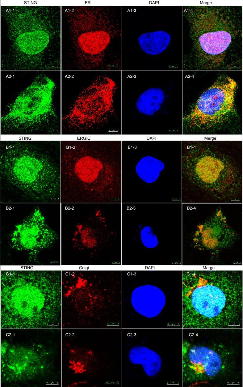
Submitted references Mitochondrial DNA drives noncanonical inflammation activation via cGAS-STING signaling pathway in retinal microvascular endothelial cells.
Guo Y, Gu R, Gan D, Hu F, Li G, Xu G
Cell communication and signaling : CCS 2020 Oct 28;18(1):172
Cell communication and signaling : CCS 2020 Oct 28;18(1):172
No comments: Submit comment
Supportive validation
- Submitted by
- Invitrogen Antibodies (provider)
- Main image
- Experimental details
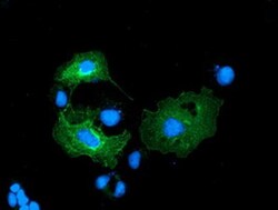
- Immunofluorescent analysis of LMAN1 in COS7 cells. Cells were transfected with a plasmid overexpressing LMAN1 and probed with a LMAN1 monoclonal antibody (Product # MA5-25345).

- Submitted by
- Invitrogen Antibodies (provider)
- Main image

- Experimental details
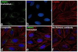
- Immunofluorescence analysis of LMAN1 was performed using 70% confluent log phase HeLa cells treated with Brefeldin A (5 µg/mL, 15 minutes). The cells were fixed with 4% paraformaldehyde for 10 minutes, permeabilized with 0.1% Triton™ X-100 for 15 minutes, and blocked with 2% BSA for 1 hour at room temperature. The cells were labeled with LMAN1 Monoclonal Antibody (OTI1A8) (Product # MA5-25345) at 1:100 dilution in 0.1% BSA, incubated at 4 degree Celsius overnight and then labeled with Goat anti-Mouse IgG (H+L) Superclonal™ Recombinant Secondary Antibody, Alexa Fluor® 488 conjugate (Product # A28175) at a dilution of 1:2000 for 45 minutes at room temperature (Panel a: green). Nuclei (Panel b: blue) were stained with ProLong™ Diamond Antifade Mountant with DAPI (Product # P36962). F-actin (Panel c: red) was stained with Rhodamine Phalloidin (Product # R415, 1:300). Panel d represents the merged image showing increase in cytoplasm expression of LMAN1. Panel e represents untreated cells, showing lesser expression of LMAN1. Panel f represents control cells with no primary antibody to assess background. The images were captured at 60X magnification.

- Submitted by
- Invitrogen Antibodies (provider)
- Main image
- Experimental details
- Immunofluorescence analysis of LMAN1 was performed using 70% confluent log phase HeLa cells treated with Brefeldin A (5 µg/mL, 15 minutes). The cells were fixed with 4% paraformaldehyde for 10 minutes, permeabilized with 0.1% Triton™ X-100 for 15 minutes, and blocked with 2% BSA for 1 hour at room temperature. The cells were labeled with LMAN1 Monoclonal Antibody (OTI1A8) (Product # MA5-25345) at 1:100 dilution in 0.1% BSA, incubated at 4 degree Celsius overnight and then labeled with Goat anti-Mouse IgG (H+L) Superclonal™ Recombinant Secondary Antibody, Alexa Fluor® 488 conjugate (Product # A28175) at a dilution of 1:2000 for 45 minutes at room temperature (Panel a: green). Nuclei (Panel b: blue) were stained with ProLong™ Diamond Antifade Mountant with DAPI (Product # P36962). F-actin (Panel c: red) was stained with Rhodamine Phalloidin (Product # R415, 1:300). Panel d represents the merged image showing increase in cytoplasm expression of LMAN1. Panel e represents untreated cells, showing lesser expression of LMAN1. Panel f represents control cells with no primary antibody to assess background. The images were captured at 60X magnification.

Supportive validation
- Submitted by
- Invitrogen Antibodies (provider)
- Main image

- Experimental details
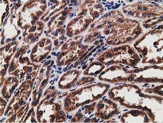
- Immunohistochemistry was performed on paraffin-embedded human kidney tissue. To expose target proteins, 10mM citric buffer, pH6.0, 100°C for 10min was used. Following antigen retrieval, tissues were probed with a LMAN1 monoclonal antibody (Product # MA5-25345).
- Submitted by
- Invitrogen Antibodies (provider)
- Main image
- Experimental details
- Immunohistochemistry was performed on paraffin-embedded carcinoma of human kidney tissue. To expose target proteins, 10mM citric buffer, pH6.0, 100°C for 10min was used. Following antigen retrieval, tissues were probed with a LMAN1 monoclonal antibody (Product # MA5-25345).

- Submitted by
- Invitrogen Antibodies (provider)
- Main image

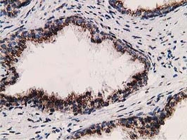
- Experimental details
- Immunohistochemistry was performed on paraffin-embedded human prostate tissue. To expose target proteins, 10mM citric buffer, pH6.0, 100°C for 10min was used. Following antigen retrieval, tissues were probed with a LMAN1 monoclonal antibody (Product # MA5-25345).
- Submitted by
- Invitrogen Antibodies (provider)
- Main image

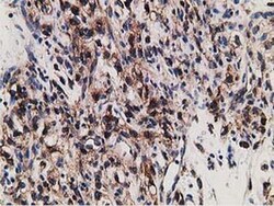
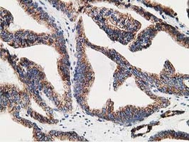
- Experimental details
- Immunohistochemistry was performed on paraffin-embedded carcinoma of human prostate tissue. To expose target proteins, 10mM citric buffer, pH6.0, 100°C for 10min was used. Following antigen retrieval, tissues were probed with a LMAN1 monoclonal antibody (Product # MA5-25345).
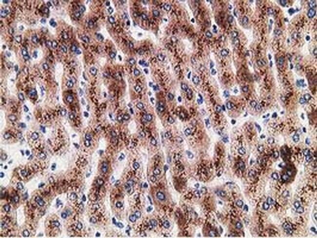
- Submitted by
- Invitrogen Antibodies (provider)
- Main image
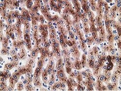
- Experimental details
- Immunohistochemistry was performed on paraffin-embedded human liver tissue. To expose target proteins, 10mM citric buffer, pH6.0, 100°C for 10min was used. Following antigen retrieval, tissues were probed with a LMAN1 monoclonal antibody (Product # MA5-25345).
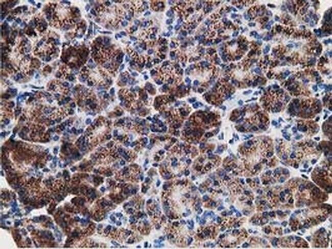
- Submitted by
- Invitrogen Antibodies (provider)
- Main image

- Experimental details
- Immunohistochemistry was performed on paraffin-embedded human pancreas tissue. To expose target proteins, 10mM citric buffer, pH6.0, 100°C for 10min was used. Following antigen retrieval, tissues were probed with a LMAN1 monoclonal antibody (Product # MA5-25345).
Supportive validation
- Submitted by
- Invitrogen Antibodies (provider)
- Main image
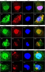
- Experimental details
- Fig. 4 Immunofluorescence of ER, ERGIC and Golgi and STING after mtDNA stimulation of RMECs. Figure A1-C1: normal control groups; Figure A2-C2: mtDNA stimulation groups. Green was STING and blue was DAPI. Red was ER, ERGIC and Golgi in figure A2-2, B2-2 and C2-2, respectively. In normal control group, STING was dispersive and co-localization with ER maker. In mtDNA-stimulated group, STING was aggregated specks formed in the perinuclear region and partly co-localization in ERGIC and Golgi makers