 Explore
 Validate
 Learn
LearnRQ4585
antibody from NSJ Bioreagents
Targeting: LMAN1
ERGIC-53, ERGIC53, F5F8D, FMFD1, gp58, MCFD1, MR60
 Western blot
 ELISA
ELISAAntibody data
- Antibody Data
- Antigen structure
- References [0]
- Comments [0]
- Validations
- Western blot [3]
- Immunocytochemistry [1]
- Immunohistochemistry [8]
Submit
Validation data
Reference
Comment
Report error
- Product number
- RQ4585 - Provider product page

- Provider
- NSJ Bioreagents
- Product name
- LMAN1 Antibody / ERGIC-53
- Antibody type
- Polyclonal
- Description
- This highly specific LMAN1 antibody is suitable for use in Western blot/Immunohistochemistry/Immunofluorescence/Flow cytometry/Direct ELISA applications with human, mouse and rat samples.
- Reactivity
- Human, Mouse, Rat
- Host
- Rabbit
- Conjugate
- Unconjugated
- Vial size
- 100 ug
- Concentration
- 0.5mg/ml if reconstituted with 0.2ml sterile DI water
- Storage
- After reconstitution, the LMAN1 antibody can be stored for up to one month at 4oC. For long-term, aliquot and store at -20oC. Avoid repeated freezing and thawing.
No comments: Submit comment
Supportive validation
- Submitted by
- NSJ Bioreagents (provider)
- Main image

- Experimental details
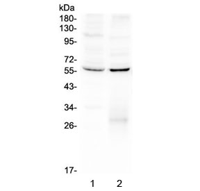
- Western blot testing of human 1) HeLa and 2) A431 lysate with LMAN1 antibody at 0.5ug/ml. Predicted molecular weight ~53 kDa.
- Submitted by
- NSJ Bioreagents (provider)
- Main image
- Experimental details
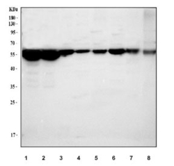
- Western blot testing of 1) human A431, 2) human HepG2, 3) rat liver, 4) rat heart, 5) rat C6, 6) mouse liver, 7) mouse heart and 8) mouse NIH 3T3 cell lysate with LMAN1 antibody. Predicted molecular weight ~53 kDa.

- Submitted by
- NSJ Bioreagents (provider)
- Main image
- Experimental details
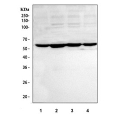
- Western blot testing of human 1) A549, 2) HepG2, 3) HeLa and 4) 293T cell lysate with LMAN1 antibody. Predicted molecular weight ~53 kDa.

Supportive validation
- Submitted by
- NSJ Bioreagents (provider)
- Main image
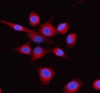
- Experimental details
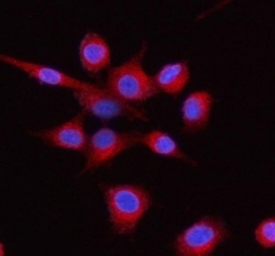
- Immunofluorescent staining of FFPE human A549 cells with LMAN1 antibody (red) and DAPI nuclear stain (blue). HIER: steam section in pH6 citrate buffer for 20 min.
Supportive validation
- Submitted by
- NSJ Bioreagents (provider)
- Main image
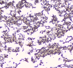
- Experimental details
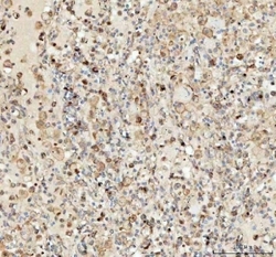
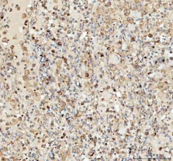
- IHC staining of FFPE human glioma with LMAN1 antibody at 1ug/ml. HIER: boil tissue sections in pH6, 10mM citrate buffer, for 10-20 min followed by cooling at RT for 20 min.
- Submitted by
- NSJ Bioreagents (provider)
- Main image
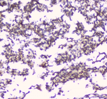
- Experimental details
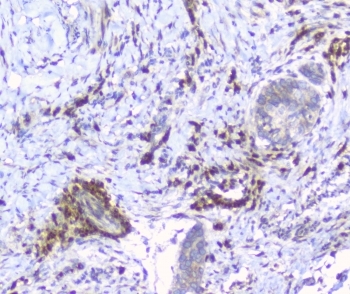
- IHC staining of FFPE human esophagus squama cancer with LMAN1 antibody at 1ug/ml. HIER: boil tissue sections in pH6, 10mM citrate buffer, for 10-20 min followed by cooling at RT for 20 min.
- Submitted by
- NSJ Bioreagents (provider)
- Main image
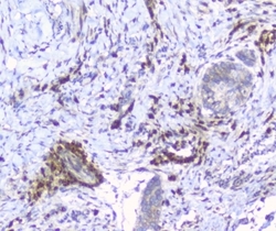
- Experimental details
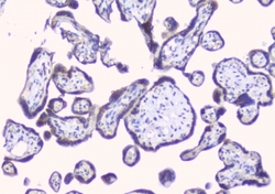
- IHC staining of FFPE human placenta with LMAN1 antibody at 1ug/ml. HIER: boil tissue sections in pH6, 10mM citrate buffer, for 10-20 min followed by cooling at RT for 20 min.

- Submitted by
- NSJ Bioreagents (provider)
- Main image

- Experimental details
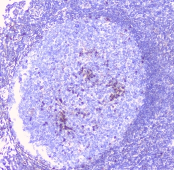
- IHC staining of FFPE human tonsil with LMAN1 antibody at 1ug/ml. HIER: boil tissue sections in pH6, 10mM citrate buffer, for 10-20 min followed by cooling at RT for 20 min.
- Submitted by
- NSJ Bioreagents (provider)
- Main image
- Experimental details
- IHC staining of FFPE human lung cancer tissue with LMAN1 antibody. HIER: boil tissue sections in pH8 EDTA buffer, for 10-20 min followed by cooling at RT for 20 min.
- Submitted by
- NSJ Bioreagents (provider)
- Main image
- Experimental details
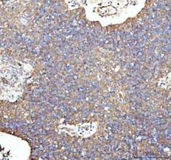
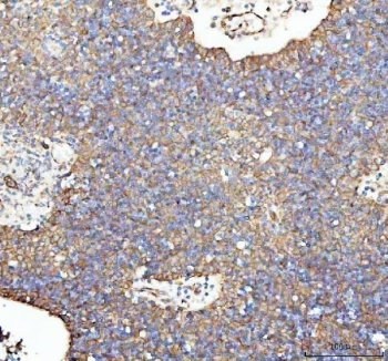
- IHC staining of FFPE human spleen tissue with LMAN1 antibody. HIER: boil tissue sections in pH8 EDTA buffer, for 10-20 min followed by cooling at RT for 20 min.
- Submitted by
- NSJ Bioreagents (provider)
- Main image

- Experimental details
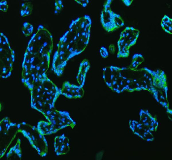
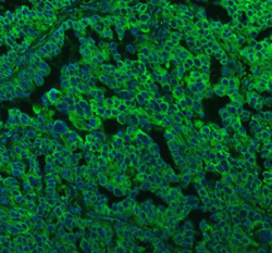
- Immunofluorescent staining of FFPE human placental tissue with LMAN1 antibody (green) and DAPI nuclear stain (blue). HIER: steam section in pH8 EDTA buffer for 20 min.
- Submitted by
- NSJ Bioreagents (provider)
- Main image
- Experimental details
- Immunofluorescent staining of FFPE human ovarian cancer tissue with LMAN1 antibody (green) and DAPI nuclear stain (blue). HIER: steam section in pH8 EDTA buffer for 20 min.
