 Explore
 Validate
 Learn
LearnPA1-074
antibody from Invitrogen Antibodies
Targeting: LMAN1
ERGIC-53, ERGIC53, F5F8D, FMFD1, gp58, MCFD1, MR60
 Western blot
Antibody data
- Antibody Data
- Antigen structure
- References [2]
- Comments [0]
- Validations
- Western blot [3]
- Immunocytochemistry [7]
- Other assay [1]
Submit
Validation data
Reference
Comment
Report error
- Product number
- PA1-074 - Provider product page

- Provider
- Invitrogen Antibodies
- Product name
- LMAN1 Polyclonal Antibody
- Antibody type
- Polyclonal
- Antigen
- Synthetic peptide
- Description
- PA1-074 detects ERGIC-53 in rat, mouse and human samples. PA1-074 has been successfully used in Western blot and immunofluorescence procedures. By Western blot, this antibody detects a ~57.5 kDa protein representing ERGIC-53 in rat brain samples. The PA1-074 immunogen is a synthetic peptide corresponding to residues F(159) D S F D N D G K K N N P A I(173) of rat ERGIC-53. This sequence is conserved in mouse and human. The PA1-074 immunizing peptide (Cat. # PEP-277) is available for use in neutralization and control experiments.
- Reactivity
- Human, Mouse, Rat
- Host
- Rabbit
- Isotype
- IgG
- Vial size
- 100 µg
- Concentration
- 1 mg/mL
- Storage
- -20° C, Avoid Freeze/Thaw Cycles
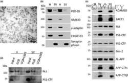
Submitted references ADAM10 and BACE1 are localized to synaptic vesicles.
Synaptic and endosomal localization of active gamma-secretase in rat brain.
Lundgren JL, Ahmed S, Schedin-Weiss S, Gouras GK, Winblad B, Tjernberg LO, Frykman S
Journal of neurochemistry 2015 Nov;135(3):606-15
Journal of neurochemistry 2015 Nov;135(3):606-15
Synaptic and endosomal localization of active gamma-secretase in rat brain.
Frykman S, Hur JY, Frånberg J, Aoki M, Winblad B, Nahalkova J, Behbahani H, Tjernberg LO
PloS one 2010 Jan 28;5(1):e8948
PloS one 2010 Jan 28;5(1):e8948
No comments: Submit comment
Supportive validation
- Submitted by
- Invitrogen Antibodies (provider)
- Main image
- Experimental details
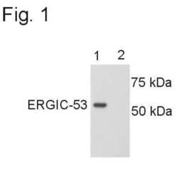
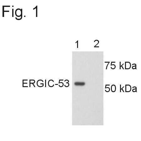
- Western blot detection of ERGIC-53 from rat brain using Product # PA1-074. Lane 1 shows the rat brain sample and lane 2 shows inhibition of ERGIC-53 due to the addition of the peptide.
- Submitted by
- Invitrogen Antibodies (provider)
- Main image
- Experimental details
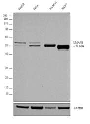
- Western blot analysis was performed on whole cell extract (30 µg lysate) of HepG2 (Lane 1), HeLa (Lane 2), PANC-1 (Lane 3), and MCF7 (Lane 4). The blot was probed with Anti-LMAN1 Polyclonal Antibody (Product # PA1-074, 1:2000 dilution) and detected by chemiluminescence using Goat anti-Rabbit IgG (H+L) Superclonal™ Secondary Antibody, HRP conjugate (Product # A27036, 0.25 µg/ml, 1:4000 dilution). A 53 kDa band corresponding to LMAN1 was detected in cell lines tested.

- Submitted by
- Invitrogen Antibodies (provider)
- Main image
- Experimental details
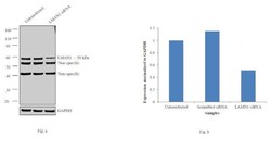
- Knockdown of LMAN1 was achieved by transfecting HeLa cells with LMAN1 specific siRNAs (Silencer® select Product # s138227, s138226). Western blot analysis (Fig. a) was performed using whole cell extracts from the LMAN1 knockdown cells (lane 3), non-specific scrambled siRNA transfected cells (lane 2) and untransfected cells (lane 1). The blots were probed with LMAN1 Polyclonal Antibody (Product # PA1-074, 1:2000 dilution) and Goat anti-Rabbit IgG (H+L) Superclonal™ Secondary Antibody, HRP conjugate (Product # A27036, 0.25 µg/ml, 1:4000 dilution). Densitometric analysis of this western blot is shown in histogram (Fig. b). Decrease in signal upon siRNA mediated knock down confirms that antibody is specific to LMAN1.

Supportive validation
- Submitted by
- Invitrogen Antibodies (provider)
- Main image
- Experimental details
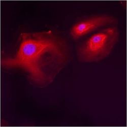
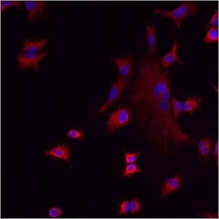
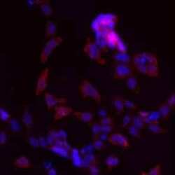
- Immunofluorescent analysis of ERGIC-53 using anti-ERGIC-53 polyclonal antibody (Product # PA1-074) shows staining in HMVEC Cells.

- Submitted by
- Invitrogen Antibodies (provider)
- Main image
- Experimental details
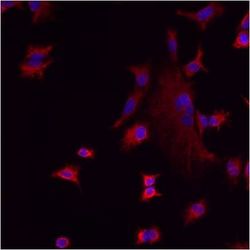
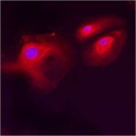
- Immunofluorescent analysis of ERGIC-53 using anti-ERGIC-53 polyclonal antibody (Product # PA1-074) shows staining in A549 Cells.
- Submitted by
- Invitrogen Antibodies (provider)
- Main image
- Experimental details
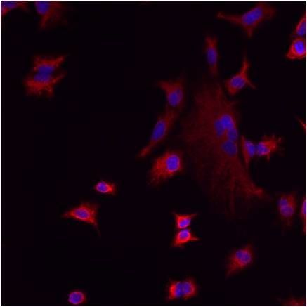
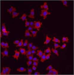
- Immunofluorescent analysis of ERGIC-53 using anti-ERGIC-53 polyclonal antibody (Product # PA1-074) shows staining in A549 Cells.
- Submitted by
- Invitrogen Antibodies (provider)
- Main image

- Experimental details
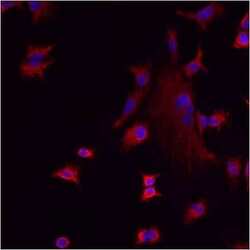
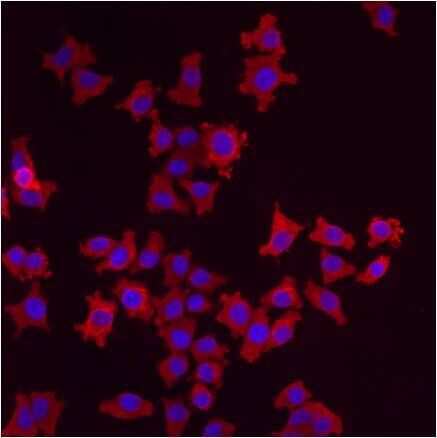
- Immunofluorescent analysis of ERGIC-53 using anti-ERGIC-53 polyclonal antibody (Product # PA1-074) shows staining in HMVEC Cells.
- Submitted by
- Invitrogen Antibodies (provider)
- Main image
- Experimental details
- Immunofluorescent analysis of ERGIC-53 using anti-ERGIC-53 polyclonal antibody (Product # PA1-074) shows staining in p19 Cells.
- Submitted by
- Invitrogen Antibodies (provider)
- Main image
- Experimental details
- Immunofluorescent analysis of ERGIC-53 using anti-ERGIC-53 polyclonal antibody (Product # PA1-074) shows staining in p19 Cells.

- Submitted by
- Invitrogen Antibodies (provider)
- Main image
- Experimental details
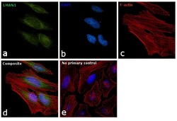
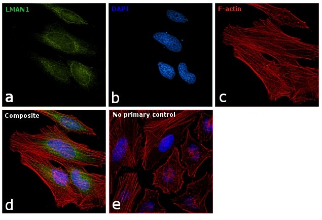
- Immunofluorescence analysis of LMAN1 was performed using 70% confluent log phase HeLa cells. The cells were fixed with 4% paraformaldehyde for 10 minutes, permeabilized with 0.1% Triton™ X-100 for 15 minutes, and blocked with 1% BSA for 1 hour at room temperature. The cells were labeled with LMAN1 Polyclonal Antibody (Product # PA1-074) at 1:200 dilution in 0.1% BSA, incubated at 4 degree Celsius overnight and then labeled with Goat anti-Rabbit IgG (H+L) Superclonal™ Secondary Antibody, Alexa Fluor® 488 conjugate (Product # A27034) at a dilution of 1:2000 for 45 minutes at room temperature (Panel a: green). Nuclei (Panel b: blue) were stained with SlowFade® Gold Antifade Mountant with DAPI (Product # S36938). F-actin (Panel c: red) was stained with Rhodamine Phalloidin (Product # R415, 1:300). Panel d represents the merged image showing endoplasmic reticulum and golgi localization. Panel e represents control cells with no primary antibody to assess background. The images were captured at 60X magnification.
Supportive validation
- Submitted by
- Invitrogen Antibodies (provider)
- Main image
- Experimental details
- NULL
