Explore
 Validate
 Learn
Learn Western blot
 Immunocytochemistry
Antibody data
- Antibody Data
- Antigen structure
- References [7]
- Comments [0]
- Validations
- Immunocytochemistry [1]
- Immunohistochemistry [3]
- Other assay [1]
Submit
Validation data
Reference
Comment
Report error
- Product number
- MA5-13330 - Provider product page

- Provider
- Invitrogen Antibodies
- Product name
- Anti-CDK6 Monoclonal Antibody (K6.83 (DCS-83)), Biotin
- Antibody type
- Monoclonal
- Antigen
- Recombinant full-length protein
- Description
- MA5-13338 targets Cdk6 in IF, IHC (F), IP, and WB applications and shows reactivity with Human, mouse, and Rat samples. The MA5-13338 immunogen is purified recombinant cdk6 protein.
- Reactivity
- Human, Mouse, Rat
- Host
- Mouse
- Conjugate
- Biotin
- Isotype
- IgG
- Antibody clone number
- K6.83 (DCS-83)
- Vial size
- 500 µL
- Concentration
- 0.2 mg/ml
- Storage
- 4° C
Submitted references Menthol inhibits the proliferation and motility of prostate cancer DU145 cells.
Death effector domain-containing protein (DEDD) is required for uterine decidualization during early pregnancy in mice.
Differential regulation of cyclin-dependent kinase 4 (CDK4) and CDK6, evidence that CDK4 might not be activated by CDK7, and design of a CDK6 activating mutation.
Regulated activating Thr172 phosphorylation of cyclin-dependent kinase 4(CDK4): its relationship with cyclins and CDK "inhibitors".
Differential utilization of cyclin D1 and cyclin D3 in the distinct mitogenic stimulations by growth factors and TSH of human thyrocytes in primary culture.
Growth inhibition of human hepatoma cells by acyclic retinoid is associated with induction of p21(CIP1) and inhibition of expression of cyclin D1.
v-Jun overrides the mitogen dependence of S-phase entry by deregulating retinoblastoma protein phosphorylation and E2F-pocket protein interactions as a consequence of enhanced cyclin E-cdk2 catalytic activity.
Wang Y, Wang X, Yang Z, Zhu G, Chen D, Meng Z
Pathology oncology research : POR 2012 Oct;18(4):903-10
Pathology oncology research : POR 2012 Oct;18(4):903-10
Death effector domain-containing protein (DEDD) is required for uterine decidualization during early pregnancy in mice.
Mori M, Kitazume M, Ose R, Kurokawa J, Koga K, Osuga Y, Arai S, Miyazaki T
The Journal of clinical investigation 2011 Jan;121(1):318-27
The Journal of clinical investigation 2011 Jan;121(1):318-27
Differential regulation of cyclin-dependent kinase 4 (CDK4) and CDK6, evidence that CDK4 might not be activated by CDK7, and design of a CDK6 activating mutation.
Bockstaele L, Bisteau X, Paternot S, Roger PP
Molecular and cellular biology 2009 Aug;29(15):4188-200
Molecular and cellular biology 2009 Aug;29(15):4188-200
Regulated activating Thr172 phosphorylation of cyclin-dependent kinase 4(CDK4): its relationship with cyclins and CDK "inhibitors".
Bockstaele L, Kooken H, Libert F, Paternot S, Dumont JE, de Launoit Y, Roger PP, Coulonval K
Molecular and cellular biology 2006 Jul;26(13):5070-85
Molecular and cellular biology 2006 Jul;26(13):5070-85
Differential utilization of cyclin D1 and cyclin D3 in the distinct mitogenic stimulations by growth factors and TSH of human thyrocytes in primary culture.
Paternot S, Dumont JE, Roger PP
Molecular endocrinology (Baltimore, Md.) 2006 Dec;20(12):3279-92
Molecular endocrinology (Baltimore, Md.) 2006 Dec;20(12):3279-92
Growth inhibition of human hepatoma cells by acyclic retinoid is associated with induction of p21(CIP1) and inhibition of expression of cyclin D1.
Suzui M, Masuda M, Lim JT, Albanese C, Pestell RG, Weinstein IB
Cancer research 2002 Jul 15;62(14):3997-4006
Cancer research 2002 Jul 15;62(14):3997-4006
v-Jun overrides the mitogen dependence of S-phase entry by deregulating retinoblastoma protein phosphorylation and E2F-pocket protein interactions as a consequence of enhanced cyclin E-cdk2 catalytic activity.
Clark W, Black EJ, MacLaren A, Kruse U, LaThangue N, Vogt PK, Gillespie DA
Molecular and cellular biology 2000 Apr;20(7):2529-42
Molecular and cellular biology 2000 Apr;20(7):2529-42
No comments: Submit comment
Supportive validation
- Submitted by
- Invitrogen Antibodies (provider)
- Main image
- Experimental details
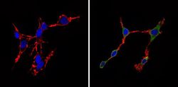
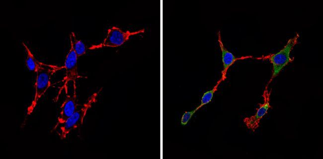
- Immunofluorescent analysis of Cdk6 (green) showing staining in the cytoplasm of C6 cells (right) compared to a negative control without primary antibody (left). Formalin-fixed cells were permeabilized with 0.1% Triton X-100 in TBS for 5-10 minutes and blocked with 3% BSA-PBS for 30 minutes at room temperature. Cells were probed with a Cdk6 monoclonal antibody (Product # MA5-13333) in 3% BSA-PBS at a dilution of 1:20 and incubated overnight at 4 ºC in a humidified chamber. Cells were washed with PBST and incubated with a DyLight-conjugated secondary antibody in PBS at room temperature in the dark. F-actin (red) was stained with a flourescent red phalloidin and nuclei (blue) were stained with Hoechst or DAPI. Images were taken at a magnification of 60x.
- Conjugate
- Biotin
Supportive validation
- Submitted by
- Invitrogen Antibodies (provider)
- Main image

- Experimental details
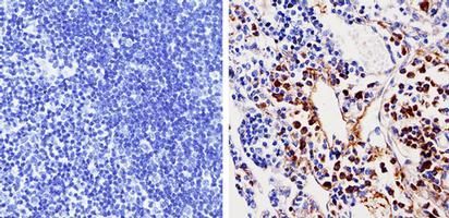
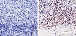
- Immunohistochemistry analysis of Cdk6 showing staining in the nucleus and cytoplasm of paraffin-treated mouse lymph node tissue (right) compared with a negative control in the absence of primary antibody (left). To expose target proteins, antigen retrieval was performed using 10mM sodium citrate (pH 6.0), microwaved for 8-15 min. Following antigen retrieval, tissues were blocked in 3% H2O2-methanol for 15 min at room temperature, washed with ddH2O and PBS, and then probed with a Cdk6 monoclonal antibody (Product # MA5-13333) diluted by 3% BSA-PBS at a dilution of 1:100 overnight at 4°C in a humidified chamber. Tissues were washed extensively in PBST and detection was performed using an HRP-conjugated secondary antibody followed by colorimetric detection using a DAB kit. Tissues were counterstained with hematoxylin and dehydrated with ethanol and xylene to prep for mounting.
- Conjugate
- Biotin
- Submitted by
- Invitrogen Antibodies (provider)
- Main image
- Experimental details
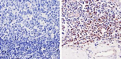
- Immunohistochemistry analysis of Cdk6 showing staining in the nucleus and cytoplasm of paraffin-treated human tonsil tissue (right) compared with a negative control in the absence of primary antibody (left). To expose target proteins, antigen retrieval was performed using 10mM sodium citrate (pH 6.0), microwaved for 8-15 min. Following antigen retrieval, tissues were blocked in 3% H2O2-methanol for 15 min at room temperature, washed with ddH2O and PBS, and then probed with a Cdk6 monoclonal antibody (Product # MA5-13333) diluted by 3% BSA-PBS at a dilution of 1:20 overnight at 4°C in a humidified chamber. Tissues were washed extensively in PBST and detection was performed using an HRP-conjugated secondary antibody followed by colorimetric detection using a DAB kit. Tissues were counterstained with hematoxylin and dehydrated with ethanol and xylene to prep for mounting.
- Conjugate
- Biotin
- Submitted by
- Invitrogen Antibodies (provider)
- Main image
- Experimental details
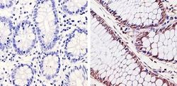
- Immunohistochemistry analysis of Cdk6 showing staining in the nucleus and cytoplasm of paraffin-treated human colon carcinoma (right) compared with a negative control in the absence of primary antibody (left). To expose target proteins, antigen retrieval was performed using 10mM sodium citrate (pH 6.0), microwaved for 8-15 min. Following antigen retrieval, tissues were blocked in 3% H2O2-methanol for 15 min at room temperature, washed with ddH2O and PBS, and then probed with a Cdk6 monoclonal antibody (Product # MA5-13333) diluted by 3% BSA-PBS at a dilution of 1:20 overnight at 4°C in a humidified chamber. Tissues were washed extensively in PBST and detection was performed using an HRP-conjugated secondary antibody followed by colorimetric detection using a DAB kit. Tissues were counterstained with hematoxylin and dehydrated with ethanol and xylene to prep for mounting.
- Conjugate
- Biotin

Supportive validation
- Submitted by
- Invitrogen Antibodies (provider)
- Main image
- Experimental details
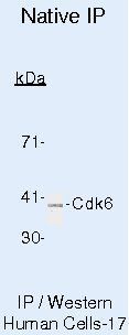
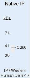
- Immunoprecipitation of Cdk6 using Cdk6 Monoclonal Antibody (Product # MA5-13330) on Native Human LS174T Cells.
- Conjugate
- Biotin