Explore
 Validate
 Learn
Learn Western blot
 Immunocytochemistry
Antibody data
- Antibody Data
- Antigen structure
- References [1]
- Comments [0]
- Validations
- Immunocytochemistry [1]
- Immunohistochemistry [1]
- Flow cytometry [2]
- Other assay [3]
Submit
Validation data
Reference
Comment
Report error
- Product number
- MA5-23833 - Provider product page

- Provider
- Invitrogen Antibodies
- Product name
- MIP-3 beta Monoclonal Antibody (54909)
- Antibody type
- Monoclonal
- Antigen
- Recombinant full-length protein
- Reactivity
- Human
- Host
- Mouse
- Isotype
- IgG
- Antibody clone number
- 54909
- Vial size
- 100 μg
- Concentration
- 1.0 mg/mL
- Storage
- -20°C, Avoid Freeze/Thaw Cycles
Submitted references CALR-TLR4 Complex Inhibits Non-Small Cell Lung Cancer Progression by Regulating the Migration and Maturation of Dendritic Cells.
Chen R, Huang M, Yang X, Chen XH, Shi MY, Li ZF, Chen ZN, Wang K
Frontiers in oncology 2021;11:743050
Frontiers in oncology 2021;11:743050
No comments: Submit comment
Supportive validation
- Submitted by
- Invitrogen Antibodies (provider)
- Main image
- Experimental details
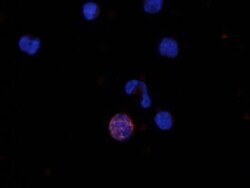
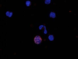
- Immunocytochemistry analysis of MIP-3 beta in immersion fixed human peripheral blood mononuclear cells (PBMCs). Samples were incubated in MIP-3 beta monoclonal antibody (Product # MA5-23833) using a dilution of 10 µg/mL for 3 hours at room temperature followed by NorthernLights™ 557-conjugated Anti-Mouse IgG Secondary Antibody (red) and counterstained with DAPI (blue).
Supportive validation
- Submitted by
- Invitrogen Antibodies (provider)
- Main image

- Experimental details
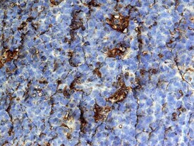
- Immunohistochemical analysis of MIP-3 beta in immersion fixed paraffin-embedded sections of human tonsil. Samples were incubated in MIP-3 beta monoclonal antibody (Product # MA5-23833) using a dilution of 25 µg/mL overnight at 4 °C. Tissue was stained with the Anti-Mouse HRP-DAB Cell & Tissue Staining Kit (brown) and counterstained with hematoxylin (blue).
Supportive validation
- Submitted by
- Invitrogen Antibodies (provider)
- Main image

- Experimental details
- Flow cytometry of MIP-3 beta in Human monocyte-derived dendritic cells. Samples were incubated in MIP-3 beta monoclonal antibody (Product # MA5-23833) or isotype control antibody followed by Phycoerythrin-conjugated Anti-Mouse IgG Secondary Antibody. To facilitate intracellular staining, cells were fixed with Flow Cytometry Fixation Buffer and permeabilized with Flow Cytometry Permeabilization/Wash Buffer.

Supportive validation
- Submitted by
- Invitrogen Antibodies (provider)
- Main image
- Experimental details
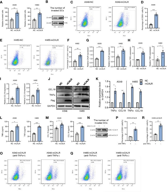
- Figure 2 mCALR facilitates the migration and maturation of DCs by increasing the secretion of TNFalpha and CCL19. (A) The effect of cell supernatants (NC and mCALR) on DC migration was evaluated by migration assay; ** P < 0.01. (B) The number of invaded DCs in both NC and mCALR groups was quantified by western blot using anti-beta-actin antibody. (C-F) The effect of cell supernatants (NC and mCALR) on DC maturation was determined using flow cytometry with PE-anti-human CD83 antibody; ** P < 0.01, *** P < 0.001. (G-I) The relative mRNA expression levels of CALR, TNFalpha, and CCL19 were detected in NC and mCALR groups using real-time PCR; * P < 0.05, ** P < 0.01, *** P < 0.001. (J, K) The expression levels of TNFalpha and CCL19 were investigated in NC and mCALR groups using western blot; * P < 0.05, ** P < 0.01. (L, M) The expression levels of TNFalpha and CCL19 were investigated in NC and mCALR groups using ELISA; * P < 0.05, *** P < 0.001. (N) The number of invaded DCs in both anti-CCL19+ and anti-CCL19- groups was quantified by western blot using anti-beta-actin antibody. (O-R) The effect of cell supernatants (anti-TNFalpha+ and anti-TNFalpha- groups) on DC maturation was determined using flow cytometry with PE-anti-human CD83 antibody; **P < 0.01.
- Submitted by
- Invitrogen Antibodies (provider)
- Main image
- Experimental details
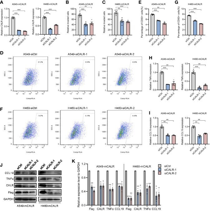
- Figure 3 mCALR silence inhibits the migration and maturation of DCs by decreasing the secretion of TNFalpha and CCL19. (A) The relative mRNA expression levels of CALR was detected in siCALR and control groups using real-time PCR; *** P < 0.001. (B, C) The effect of cell supernatants (siCtrl and siCALR) on DC migration was evaluated by migration assay; ** P < 0.01, *** P < 0.001. (D-G) The effect of cell supernatants (siCtrl and siCALR) on DC maturation was determined using flow cytometry with PE-anti-human CD83 antibody; ** P < 0.01, *** P < 0.001. (H, I) The relative mRNA expression levels of TNFalpha and CCL19 were detected in siCALR and control groups using real-time PCR; ** P < 0.01, *** P < 0.001. (J, K) The expression levels of CALR, Flag-mCALR, TNFalpha, and CCL19 were investigated in siCALR and control groups using western blot; * P < 0.05, ** P < 0.01, *** P < 0.001.
- Submitted by
- Invitrogen Antibodies (provider)
- Main image
- Experimental details
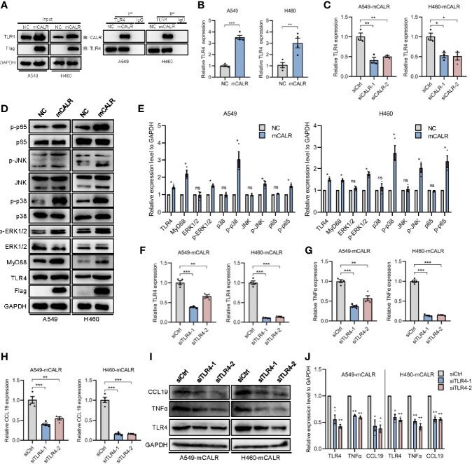
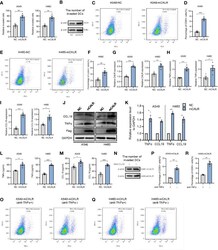
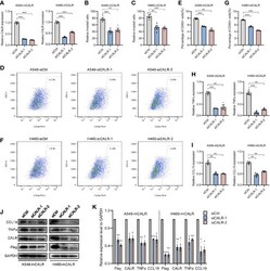
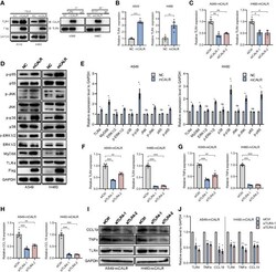
- Figure 4 mCALR enhances the secretion of TNFalpha and CCL19 via activating CALR-TLR4-MyD88 signaling. (A) The interaction between mCALR and TLR4 was assessed using co-IP assay in NC and mCALR cells, and the IgG antibody was used as a negative control. (B) The relative mRNA expression level of TLR4 was detected in NC and mCALR groups using real-time PCR; ** P < 0.01, *** P < 0.001. (C) The relative mRNA expression level of TLR4 was detected in siCALR and control groups using real-time PCR; * P < 0.05, ** P < 0.01. (D, E) The expression levels of mCALR, TLR4, MyD88, ERK1/2, p-ERK1/2, p38, p-p38, JNK, p-JNK, p65, and p-p65 were investigated in NC and mCALR groups using western blot, and the anti-Flag antibody was used to detect the mCALR protein; ns, P >0.05, * P < 0.05. (F-H) The relative mRNA expression levels of TLR4, TNFalpha, and CCL19 were detected in mCALR cells transfected with different siTLR4 using real-time PCR; ** P < 0.01, *** P < 0.001. (I, J) The expression levels of TLR4, TNFalpha, and CCL19 were investigated in mCALR cells transfected with different siTLR4 using western blot; * P < 0.05, ** P < 0.01.