Explore
 Validate
 Learn
Learn Western blot
Antibody data
- Antibody Data
- Antigen structure
- References [1]
- Comments [0]
- Validations
- Western blot [2]
- Immunocytochemistry [2]
- Flow cytometry [3]
- Other assay [1]
Submit
Validation data
Reference
Comment
Report error
- Product number
- MA5-24291 - Provider product page

- Provider
- Invitrogen Antibodies
- Product name
- EOMES Monoclonal Antibody (644730)
- Antibody type
- Monoclonal
- Antigen
- Recombinant full-length protein
- Description
- Reconstitute in sterile PBS to a final concentration of 0.5 mg/mL.
- Reactivity
- Human
- Host
- Mouse
- Isotype
- IgG
- Antibody clone number
- 644730
- Vial size
- 100 μg
- Concentration
- 0.5 mg/mL
- Storage
- -20°C, Avoid Freeze/Thaw Cycles
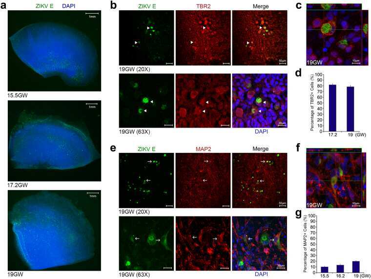
Submitted references Zika Virus Infects Intermediate Progenitor Cells and Post-mitotic Committed Neurons in Human Fetal Brain Tissues.
Lin MY, Wang YL, Wu WL, Wolseley V, Tsai MT, Radic V, Thornton ME, Grubbs BH, Chow RH, Huang IC
Scientific reports 2017 Nov 1;7(1):14883
Scientific reports 2017 Nov 1;7(1):14883
No comments: Submit comment
Supportive validation
- Submitted by
- Invitrogen Antibodies (provider)
- Main image
- Experimental details
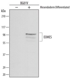
- Western blot analysis of EOMES in BG01V human embryonic stem cells untreated (-) or mesendoderm differentiated (+). Samples were incubated in EOMES monoclonal antibody (Product # MA5-24291) using a dilution of 1 µg/mL followed by a HRP-conjugated Anti-Mouse IgG secondary antibody. Specific bands were detected for EOMES at approximately 100 and 80 kDa (as indicated). This experiment was conducted under reducing conditions.

- Submitted by
- Invitrogen Antibodies (provider)
- Main image
- Experimental details
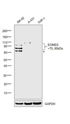
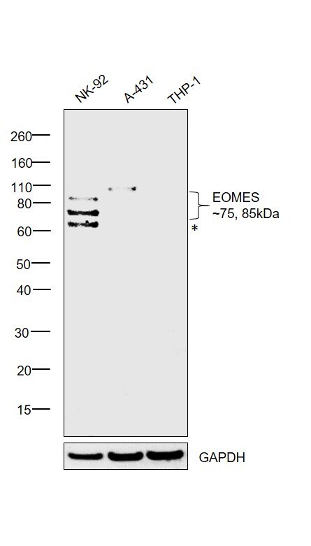
- Western blot was performed using Anti-EOMES Monoclonal Antibody (644730) (Product # MA5-24291) and 75, 85kDa bands corresponding to EOMES were observed in NK-92 but were absent in A-431 and THP-1 which are reported to be negative. Modified whole cell extracts (1% SDS) (30 µg lysate) of NK-92 (Lane 1), A-431 (Lane 2) and THP-1 (Lane 3) were electrophoresed using Novex® NuPAGE® 4-12 % Bis-Tris gel (Product # NP0322BOX). Resolved proteins were then transferred onto a nitrocellulose membrane (Product # IB23001) by iBlot® 2 Dry Blotting System (Product # IB21001). The blot was probed with the primary antibody (1µg/mL) and detected by chemiluminescence with Goat anti-Mouse IgG (H+L), Superclonal™ Recombinant Secondary Antibody, HRP (Product # A28177, 1:4000 dilution) using the iBright FL 1000 (Product # A32752). Chemiluminescent detection was performed using Novex® ECL Chemiluminescent Substrate Reagent Kit (Product # WP20005). An uncharacterized band (*) was observed at ~60kDa.
Supportive validation
- Submitted by
- Invitrogen Antibodies (provider)
- Main image
- Experimental details
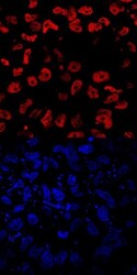
- Immunocytochemistry analysis of EOMES in immersion fixed BG01V human embryonic stem cells differentiated into mesoderm. Samples were incubated in EOMES monoclonal antibody (Product # MA5-24291) using a dilution of 10 µg/mL for 3 hours at room temperature followed by NorthernLights™ 557-conjugated Anti-Mouse IgG Secondary Antibody (red, upper panel) and counterstained with DAPI (blue, lower panel). Specific staining was localized to nuclei.

- Submitted by
- Invitrogen Antibodies (provider)
- Main image

- Experimental details
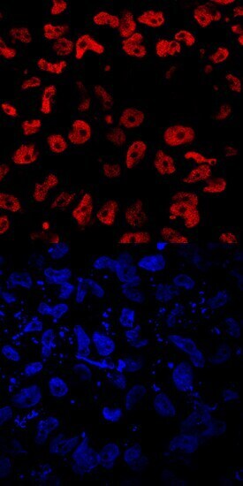
- Immunocytochemistry analysis of EOMES in immersion fixed BG01V human embryonic stem cells differentiated into mesoderm. Samples were incubated in EOMES monoclonal antibody (Product # MA5-24291) using a dilution of 10 µg/mL for 3 hours at room temperature followed by NorthernLights™ 557-conjugated Anti-Mouse IgG Secondary Antibody (red, upper panel) and counterstained with DAPI (blue, lower panel). Specific staining was localized to nuclei.
Supportive validation
- Submitted by
- Invitrogen Antibodies (provider)
- Main image

- Experimental details
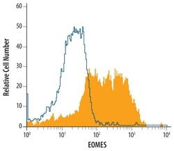
- Flow cytometric analysis of BG01V human embryonic stem cells differentiated to mesendoderm were stained with mouse Anti-human EOMES Monoclonal Antibody (Product # MA5-24291) or isotype control antibodyopen histogram), followed by Allophycocyanin-conjugated Anti-mouse IgG Secondary Antibody. To facilitate intracellular staining, cells were fixed with paraformaldehyde and permeabilized with saponin.

- Submitted by
- Invitrogen Antibodies (provider)
- Main image
- Experimental details
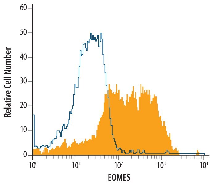
- Flow cytometry of EOMES in BG01V human embryonic stem cells differentiated to mesendoderm. Samples were incubated in EOMES monoclonal antibody (Product # MA5-24291) or isotype control antibody followed by Allophycocyanin-conjugated Anti-Mouse IgG Secondary Antibody. To facilitate intracellular staining, cells were fixed with paraformaldehyde and permeabilized with saponin.

- Submitted by
- Invitrogen Antibodies (provider)
- Main image

- Experimental details
- Flow cytometry of EOMES in BG01V human embryonic stem cells differentiated to mesendoderm. Samples were incubated in EOMES monoclonal antibody (Product # MA5-24291) or isotype control antibody followed by Allophycocyanin-conjugated Anti-Mouse IgG Secondary Antibody. To facilitate intracellular staining, cells were fixed with paraformaldehyde and permeabilized with saponin.
Supportive validation
- Submitted by
- Invitrogen Antibodies (provider)
- Main image
- Experimental details
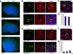
- NULL