 Explore
 Validate
 Learn
LearnAF7565
antibody from R&D Systems
Targeting: PSMA1
HC2, MGC14542, MGC14575, MGC14751, MGC1667, MGC21459, MGC22853, MGC23915, NU, PROS30
 Western blot
Antibody data
- Antibody Data
- Antigen structure
- References [0]
- Comments [0]
- Validations
- Western blot [1]
- Immunohistochemistry [1]
Submit
Validation data
Reference
Comment
Report error
- Product number
- AF7565 - Provider product page

- Provider
- R&D Systems
- Product name
- Human/Mouse/Rat PSMA1 Antibody
- Antibody type
- Polyclonal
- Description
- Immunogen affinity purified. Detects human, mouse, and rat PSMA1 in Western blots.
- Reactivity
- Human, Mouse, Rat
- Host
- Sheep
- Conjugate
- Unconjugated
- Antigen sequence
P25786- Isotype
- IgG
- Vial size
- 100 ug
- Concentration
- LYOPH
- Storage
- Use a manual defrost freezer and avoid repeated freeze-thaw cycles. 12 months from date of receipt, -20 to -70 °C as supplied. 1 month, 2 to 8 °C under sterile conditions after reconstitution. 6 months, -20 to -70 °C under sterile conditions after reconstitution.
No comments: Submit comment
Supportive validation
- Submitted by
- R&D Systems (provider)
- Main image
- Experimental details
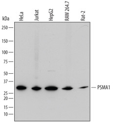
- Detection of Human, Mouse, and Rat PSMA1 by Western Blot. Western blot shows lysates of HeLa human cervical epithelial carcinoma cell line, Jurkat human acute T cell leukemia cell line, HepG2 human hepatocellular carcinoma cell line, RAW 264.7 mouse monocyte/macrophage cell line, and Rat-2 rat embryonic fibroblast cell line. PVDF membrane was probed with 0.5 µg/mL of Sheep Anti-Human/Mouse/Rat PSMA1 Antigen Affinity-purified Polyclonal Antibody (Catalog # AF7565) followed by HRP-conjugated Anti-Sheep IgG Secondary Antibody (Catalog # HAF016). A specific band was detected for PSMA1 at approximately 30 kDa (as indicated). This experiment was conducted under reducing conditions and using Immunoblot Buffer Group 1.

Supportive validation
- Submitted by
- R&D Systems (provider)
- Main image
- Experimental details
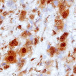
- PSMA1 in Human Breast Cancer Tissue. PSMA1 was detected in immersion fixed paraffin-embedded sections of human breast cancer tissue using Sheep Anti-Human/Mouse/Rat PSMA1 Antigen Affinity-purified Polyclonal Antibody (Catalog # AF7565) at 1.7 µg/mL overnight at 4 °C. Tissue was stained using the Anti-Sheep HRP-DAB Cell & Tissue Staining Kit (brown; Catalog # CTS019) and counterstained with hematoxylin (blue). Specific staining was localized to cytoplasm and nuclei. View our protocol for Chromogenic IHC Staining of Paraffin-embedded Tissue Sections.
