Explore
 Validate
 Learn
Learn Western blot
 ELISA
ELISAAntibody data
- Antibody Data
- Antigen structure
- References [26]
- Comments [0]
- Validations
- ELISA [2]
- Immunocytochemistry [2]
- Flow cytometry [4]
- Other assay [9]
Submit
Validation data
Reference
Comment
Report error
- Product number
- P620 - Provider product page

- Provider
- Invitrogen Antibodies
- Product name
- IL-6 Polyclonal Antibody
- Antibody type
- Polyclonal
- Antigen
- Recombinant full-length protein
- Description
- P620 detects IL-6 in WB, Flow and ELISA applications with human samples. The P620 immunogen is recombinant human IL-6. P620 detects IL-6 which has a predicted molecular weight of approximately 22 kDa. This product has been tested for endotoxins by limulus amoebocyte lysate (LAL) assay and contains an endotoxin concentration of less than or equal to 10 endotoxin units per milligram (EU/mg).
- Reactivity
- Human, Mouse
- Host
- Rabbit
- Isotype
- IgG
- Vial size
- 1 mg
- Concentration
- 1.0 mg/mL
- Storage
- -20°C
Submitted references Dihydromyricetin ameliorates osteogenic differentiation of human aortic valve interstitial cells by targeting c-KIT/interleukin-6 signaling pathway.
Design of Targeted Flurbiprofen Biomimetic Nanoparticles for Management of Arthritis: In Vitro and In Vivo Appraisal.
Luteolin Improves Cyclophosphamide-Induced Cystitis through TXNIP/NLRP3 and NF-κB Pathways.
Downregulation of APRIN expression increases cancer cell proliferation via an interleukin-6/STAT3/cyclin D axis.
β2-Adrenergic agonists attenuate organic dust-induced lung inflammation.
Redox control of the senescence regulator interleukin-1α and the secretory phenotype.
Macrophage-derived interleukin-6 up-regulates MUC1, but down-regulates MUC2 expression in the human colon cancer HT-29 cell line.
Quantification of the neutralization of cytokine biological activity by antibody: the ten-fold reduction bioassay of interleukin-6 as growth factor.
Human mast cell activation with virus-associated stimuli leads to the selective chemotaxis of natural killer cells by a CXCL8-dependent mechanism.
Combined immunogene therapy of IL-6 and IL-15 enhances anti-tumor activity through augmented NK cytotoxicity.
Interleukin-6 fused to an anti-idiotype antibody in a vaccine increases the specific humoral immune response against CA125+ (MUC-16) ovarian cancer.
Changes in macrophages in spleen and lymph nodes during acute African swine fever: expression of cytokines.
Changes in macrophages in spleen and lymph nodes during acute African swine fever: expression of cytokines.
Membrane-anchored forms of lipopolysaccharide (LPS)-binding protein do not mediate cellular responses to LPS independently of CD14.
Secretion of proinflammatory cytokines by epithelial cells in response to Chlamydia infection suggests a central role for epithelial cells in chlamydial pathogenesis.
Retrograde inflammatory signaling from neutrophils to endothelial cells by soluble interleukin-6 receptor alpha.
Interleukin-6 stimulates neutrophil production of platelet-activating factor.
Serum IL-4, IL-10 and IL-6 levels in inflammatory arthritis.
Entamoeba histolytica trophozoites induce an inflammatory cytokine response by cultured human cells through the paracrine action of cytolytically released interleukin-1 alpha.
Localization of cytokines in cholesteatoma tissue.
Localization of cytokines in cholesteatoma tissue.
Localization of cytokines in cholesteatoma tissue.
A distinct array of proinflammatory cytokines is expressed in human colon epithelial cells in response to bacterial invasion.
Sustained high levels of circulating chaperoned interleukin-6 after active specific cancer immunotherapy.
Differential expression and ligand-induced modulation of the human interleukin-6 receptor on interleukin-6-responsive cells.
Soluble IgA and IgG aggregates are catabolized by cultured rat mesangial cells and induce production of TNF-alpha and IL-6, and proliferation.
Zhang S, Fan L, Wang Y, Xu J, Shen Q, Xie J, Zeng Z, Zhou T
Frontiers in pharmacology 2022;13:932092
Frontiers in pharmacology 2022;13:932092
Design of Targeted Flurbiprofen Biomimetic Nanoparticles for Management of Arthritis: In Vitro and In Vivo Appraisal.
Mohamed HI, El-Kamel AH, Hammad GO, Heikal LA
Pharmaceutics 2022 Jan 7;14(1)
Pharmaceutics 2022 Jan 7;14(1)
Luteolin Improves Cyclophosphamide-Induced Cystitis through TXNIP/NLRP3 and NF-κB Pathways.
Zhang H, Zhao J, Lu Q, Sun B, Liu X, Yang C, Li S, Li L, Yi S, Yang Z, Xu J
Evidence-based complementary and alternative medicine : eCAM 2021;2021:1718709
Evidence-based complementary and alternative medicine : eCAM 2021;2021:1718709
Downregulation of APRIN expression increases cancer cell proliferation via an interleukin-6/STAT3/cyclin D axis.
Sohn MS, Kang M, Kang SM, Bae S
Oncology letters 2021 Jan;21(1):55
Oncology letters 2021 Jan;21(1):55
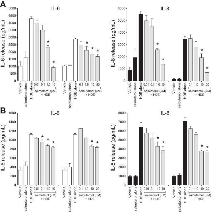
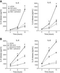
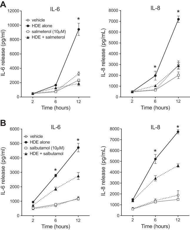
β2-Adrenergic agonists attenuate organic dust-induced lung inflammation.
Romberger DJ, Heires AJ, Nordgren TM, Poole JA, Toews ML, West WW, Wyatt TA
American journal of physiology. Lung cellular and molecular physiology 2016 Jul 1;311(1):L101-10
American journal of physiology. Lung cellular and molecular physiology 2016 Jul 1;311(1):L101-10
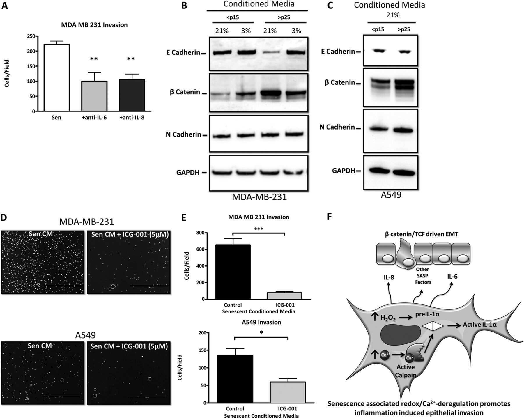
Redox control of the senescence regulator interleukin-1α and the secretory phenotype.
McCarthy DA, Clark RR, Bartling TR, Trebak M, Melendez JA
The Journal of biological chemistry 2013 Nov 8;288(45):32149-32159
The Journal of biological chemistry 2013 Nov 8;288(45):32149-32159
Macrophage-derived interleukin-6 up-regulates MUC1, but down-regulates MUC2 expression in the human colon cancer HT-29 cell line.
Li YY, Hsieh LL, Tang RP, Liao SK, Yeh KY
Cellular immunology 2009;256(1-2):19-26
Cellular immunology 2009;256(1-2):19-26
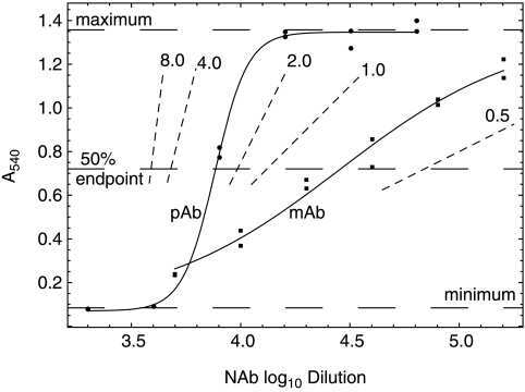
Quantification of the neutralization of cytokine biological activity by antibody: the ten-fold reduction bioassay of interleukin-6 as growth factor.
Grossberg SE, Casey M, Grossberg LD
Journal of interferon & cytokine research : the official journal of the International Society for Interferon and Cytokine Research 2009 Aug;29(8):421-6
Journal of interferon & cytokine research : the official journal of the International Society for Interferon and Cytokine Research 2009 Aug;29(8):421-6
Human mast cell activation with virus-associated stimuli leads to the selective chemotaxis of natural killer cells by a CXCL8-dependent mechanism.
Burke SM, Issekutz TB, Mohan K, Lee PW, Shmulevitz M, Marshall JS
Blood 2008 Jun 15;111(12):5467-76
Blood 2008 Jun 15;111(12):5467-76
Combined immunogene therapy of IL-6 and IL-15 enhances anti-tumor activity through augmented NK cytotoxicity.
Lin CY, Chuang TF, Liao KW, Huang YJ, Pai CC, Chu RM
Cancer letters 2008 Dec 18;272(2):285-95
Cancer letters 2008 Dec 18;272(2):285-95
Interleukin-6 fused to an anti-idiotype antibody in a vaccine increases the specific humoral immune response against CA125+ (MUC-16) ovarian cancer.
Reinartz S, Hombach A, Köhler S, Schlebusch H, Wallwiener D, Abken H, Wagner U
Cancer research 2003 Jun 15;63(12):3234-40
Cancer research 2003 Jun 15;63(12):3234-40
Changes in macrophages in spleen and lymph nodes during acute African swine fever: expression of cytokines.
Salguero FJ, Ruiz-Villamor E, Bautista MJ, Sánchez-Cordón PJ, Carrasco L, Gómez-Villamandos JC
Veterinary immunology and immunopathology 2002 Nov;90(1-2):11-22
Veterinary immunology and immunopathology 2002 Nov;90(1-2):11-22
Membrane-anchored forms of lipopolysaccharide (LPS)-binding protein do not mediate cellular responses to LPS independently of CD14.
Tapping RI, Orr SL, Lawson EM, Soldau K, Tobias PS
Journal of immunology (Baltimore, Md. : 1950) 1999 May 1;162(9):5483-9
Journal of immunology (Baltimore, Md. : 1950) 1999 May 1;162(9):5483-9
Secretion of proinflammatory cytokines by epithelial cells in response to Chlamydia infection suggests a central role for epithelial cells in chlamydial pathogenesis.
Rasmussen SJ, Eckmann L, Quayle AJ, Shen L, Zhang YX, Anderson DJ, Fierer J, Stephens RS, Kagnoff MF
The Journal of clinical investigation 1997 Jan 1;99(1):77-87
The Journal of clinical investigation 1997 Jan 1;99(1):77-87
Retrograde inflammatory signaling from neutrophils to endothelial cells by soluble interleukin-6 receptor alpha.
Modur V, Li Y, Zimmerman GA, Prescott SM, McIntyre TM
The Journal of clinical investigation 1997 Dec 1;100(11):2752-6
The Journal of clinical investigation 1997 Dec 1;100(11):2752-6
Interleukin-6 stimulates neutrophil production of platelet-activating factor.
Biffl WL, Moore EE, Moore FA, Barnett CC Jr, Silliman CC, Peterson VM
Journal of leukocyte biology 1996 Apr;59(4):569-74
Journal of leukocyte biology 1996 Apr;59(4):569-74
Serum IL-4, IL-10 and IL-6 levels in inflammatory arthritis.
Cicuttini FM, Byron KA, Maher D, Wootton AM, Muirden KD, Hamilton JA
Rheumatology international 1995;14(5):201-6
Rheumatology international 1995;14(5):201-6
Entamoeba histolytica trophozoites induce an inflammatory cytokine response by cultured human cells through the paracrine action of cytolytically released interleukin-1 alpha.
Eckmann L, Reed SL, Smith JR, Kagnoff MF
The Journal of clinical investigation 1995 Sep;96(3):1269-79
The Journal of clinical investigation 1995 Sep;96(3):1269-79
Localization of cytokines in cholesteatoma tissue.
Marenda SA, Aufdemorte TB
Otolaryngology--head and neck surgery : official journal of American Academy of Otolaryngology-Head and Neck Surgery 1995 Mar;112(3):359-68
Otolaryngology--head and neck surgery : official journal of American Academy of Otolaryngology-Head and Neck Surgery 1995 Mar;112(3):359-68
A distinct array of proinflammatory cytokines is expressed in human colon epithelial cells in response to bacterial invasion.
Jung HC, Eckmann L, Yang SK, Panja A, Fierer J, Morzycka-Wroblewska E, Kagnoff MF
The Journal of clinical investigation 1995 Jan;95(1):55-65
The Journal of clinical investigation 1995 Jan;95(1):55-65
Sustained high levels of circulating chaperoned interleukin-6 after active specific cancer immunotherapy.
May LT, Patel K, García D, Ndubuisi MI, Ferrone S, Mittelman A, Mackiewicz A, Sehgal PB
Blood 1994 Sep 15;84(6):1887-95
Blood 1994 Sep 15;84(6):1887-95
Differential expression and ligand-induced modulation of the human interleukin-6 receptor on interleukin-6-responsive cells.
Schwabe M, Zhao J, Kung HF
The Journal of biological chemistry 1994 Mar 11;269(10):7201-9
The Journal of biological chemistry 1994 Mar 11;269(10):7201-9
Soluble IgA and IgG aggregates are catabolized by cultured rat mesangial cells and induce production of TNF-alpha and IL-6, and proliferation.
Gómez-Guerrero C, López-Armada MJ, González E, Egido J
Journal of immunology (Baltimore, Md. : 1950) 1994 Dec 1;153(11):5247-55
Journal of immunology (Baltimore, Md. : 1950) 1994 Dec 1;153(11):5247-55
No comments: Submit comment
Supportive validation
- Submitted by
- Invitrogen Antibodies (provider)
- Main image

- Experimental details
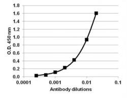
- Direct ELISA analysis of LPS-treated IL-6 was performed by coating wells of a 96-well plate with 100 µL per well of IL-6 (Product # RIL6I) diluted in carbonate/bicarbonate buffer (Product # 28382) at a concentration of 2 µg/mL overnight at 4C. Wells of the plate were washed, blocked with starting blocking buffer (Product # 37538), and incubated with 100 µL per well of a rabbit Anti-IL-6 Polyclonal Antibody (Product # P620) at a serially dilution of 1:50, 1:100, 1:250, 1:500, 1:1000, 1:2000 and 1:5000 for 90 minutes at 37C. The plate was washed, then incubated with 100 µL per well of an HRP-conjugated Goat anti-Rabbit IgG secondary antibody (Product # 65-6120) at a dilution of 1:5000 for 90 minutes at 37C. Detection was performed using 1-Step Ultra TMB substrate (Product # 34028) for 5-10 minutes at room temperature in the dark. The reaction was stopped with Stop solution (Product # N600), and absorbances were read on a spectrophotometer at 450-550 nm.

- Submitted by
- Invitrogen Antibodies (provider)
- Main image
- Experimental details
- Direct ELISA analysis of LPS-treated IL-6 was performed by coating wells of a 96-well plate with 100 µL per well of IL-6 (Product # RIL6I) diluted in carbonate/bicarbonate buffer (Product # 28382) at a concentration of 2 µg/mL overnight at 4C. Wells of the plate were washed, blocked with starting blocking buffer (Product # 37538), and incubated with 100 µL per well of a rabbit Anti-IL-6 Polyclonal Antibody (Product # P620) at a serially dilution of 1:50, 1:100, 1:250, 1:500, 1:1000, 1:2000 and 1:5000 for 90 minutes at 37C. The plate was washed, then incubated with 100 µL per well of an HRP-conjugated Goat anti-Rabbit IgG secondary antibody (Product # 65-6120) at a dilution of 1:5000 for 90 minutes at 37C. Detection was performed using 1-Step Ultra TMB substrate (Product # 34028) for 5-10 minutes at room temperature in the dark. The reaction was stopped with Stop solution (Product # N600), and absorbances were read on a spectrophotometer at 450-550 nm.

Supportive validation
- Submitted by
- Invitrogen Antibodies (provider)
- Main image

- Experimental details
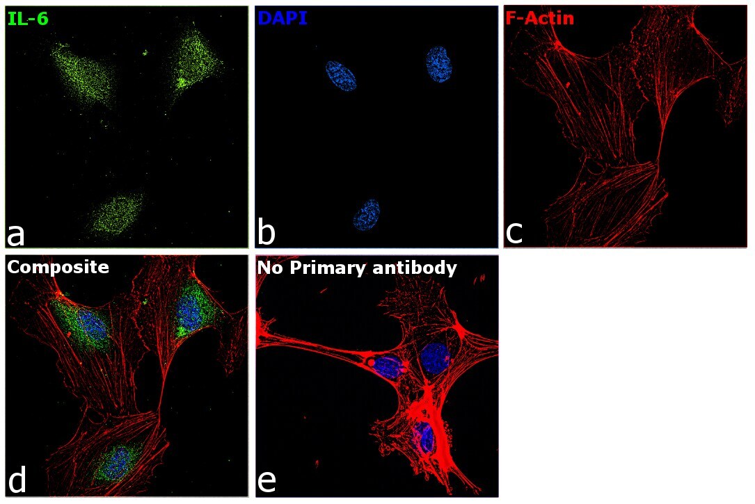
- Immunofluorescence analysis of IL-6 was performed using 70% confluent log phase HUVEC cells. The cells were fixed with 4% paraformaldehyde for 10 minutes, permeabilized with 0.1% Triton™ X-100 for 15 minutes, and blocked with 2% BSA for 45 minutes at room temperature. The cells were labeled with IL-6 Polyclonal Antibody (Product # P620) at 1:100 dilution in 0.1% BSA, incubated at 4 degree celsius overnight and then labeled with Donkey anti-Rabbit IgG (H+L) Highly Cross-Adsorbed Secondary Antibody, Alexa Fluor Plus 488 (Product # A32790), (1:2000 dilution), for 45 minutes at room temperature (Panel a: Green). Nuclei (Panel b:Blue) were stained with ProLong™ Diamond Antifade Mountant with DAPI (Product # P36962). F-actin (Panel c: Red) was stained with Rhodamine Phalloidin (Product # R415, 1:300). Panel d represents the merged image showing Predominantly Golgi Complex and Endoplasmic Reticulum localization. Panel e represents control cells with no primary antibody to assess background. The images were captured at 60X magnification.
- Submitted by
- Invitrogen Antibodies (provider)
- Main image

- Experimental details
- Immunofluorescence analysis of IL-6 was performed using 70% confluent log phase HUVEC cells. The cells were fixed with 4% paraformaldehyde for 10 minutes, permeabilized with 0.1% Triton™ X-100 for 15 minutes, and blocked with 2% BSA for 45 minutes at room temperature. The cells were labeled with IL-6 Polyclonal Antibody (Product # P620) at 1:100 dilution in 0.1% BSA, incubated at 4 degree celsius overnight and then labeled with Donkey anti-Rabbit IgG (H+L) Highly Cross-Adsorbed Secondary Antibody, Alexa Fluor Plus 488 (Product # A32790), (1:2000 dilution), for 45 minutes at room temperature (Panel a: Green). Nuclei (Panel b:Blue) were stained with ProLong™ Diamond Antifade Mountant with DAPI (Product # P36962). F-actin (Panel c: Red) was stained with Rhodamine Phalloidin (Product # R415, 1:300). Panel d represents the merged image showing Predominantly Golgi Complex and Endoplasmic Reticulum localization. Panel e represents control cells with no primary antibody to assess background. The images were captured at 60X magnification.

Supportive validation
- Submitted by
- Invitrogen Antibodies (provider)
- Main image
- Experimental details
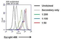
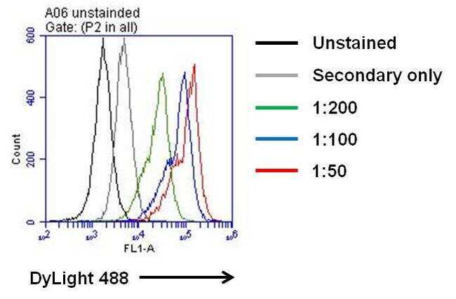
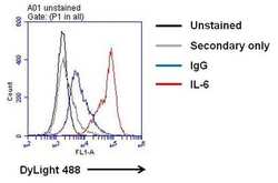
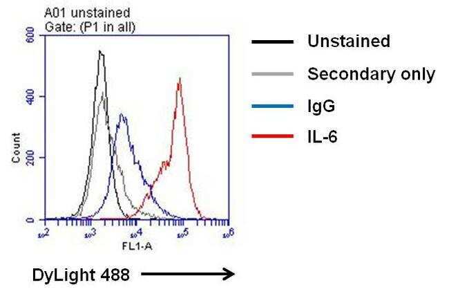
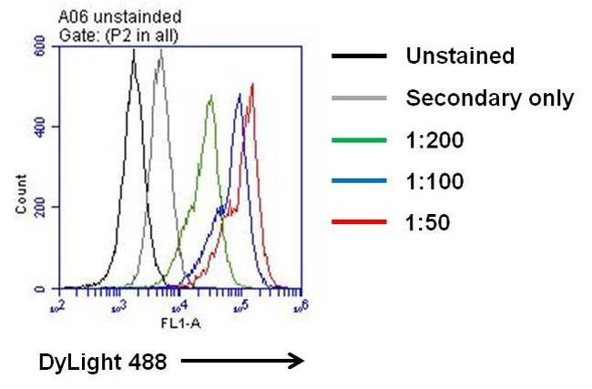
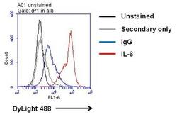
- Flow cytometry analysis of IL-6 on LPS-treated THP-1 cells. Cells were fixed with 4% formaldehyde for 30 min on ince and permeabilized with IC permeabilization buffer (Product # PB001). After incubation with blocking buffer (Product # 37525) for 30 min on ice, cells were then stained with IFN-gamma rabbit polyclonal antibody (Product # P620) at indicated dilution with IC permeabilization buffer for 30 min on ice. After washing with ice-cold IC permeabilization buffer for 3 times, the cells were stained with DyLight 488 goat anti-mouse secondary antibody (Product # 35552) for 30 min on ice. A representative 10,000 cells were acquired for each sample.
- Submitted by
- Invitrogen Antibodies (provider)
- Main image
- Experimental details
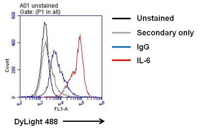
- Flow cytometry analysis of IL-6 on LPS-treated THP-1 cells. Cells were fixed with 4% formaldehyde for 30 min on ince and permeabilized with IC permeabilization buffer (Product # PB001). After incubation with blocking buffer (Product # 37525) for 30 min on ice, cells were then stained with IFN-gamma rabbit polyclonal antibody (Product # P620) or rabbit IgG control at 1:100 dilution with IC permeabilization buffer for 30 min on ice. After washing with ice-cold IC permeabilization buffer for 3 times, the cells were stained with DyLight 488 goat anti-mouse secondary antibody (Product # 35552) for 30 min on ice. A representative 10,000 cells were acquired for each sample.
- Submitted by
- Invitrogen Antibodies (provider)
- Main image
- Experimental details
- Flow cytometry analysis of IL-6 on LPS-treated THP-1 cells. Cells were fixed with 4% formaldehyde for 30 min on ince and permeabilized with IC permeabilization buffer (Product # PB001). After incubation with blocking buffer (Product # 37525) for 30 min on ice, cells were then stained with IFN-gamma rabbit polyclonal antibody (Product # P620) at indicated dilution with IC permeabilization buffer for 30 min on ice. After washing with ice-cold IC permeabilization buffer for 3 times, the cells were stained with DyLight 488 goat anti-mouse secondary antibody (Product # 35552) for 30 min on ice. A representative 10,000 cells were acquired for each sample.
- Submitted by
- Invitrogen Antibodies (provider)
- Main image
- Experimental details
- Flow cytometry analysis of IL-6 on LPS-treated THP-1 cells. Cells were fixed with 4% formaldehyde for 30 min on ince and permeabilized with IC permeabilization buffer (Product # PB001). After incubation with blocking buffer (Product # 37525) for 30 min on ice, cells were then stained with IFN-gamma rabbit polyclonal antibody (Product # P620) or rabbit IgG control at 1:100 dilution with IC permeabilization buffer for 30 min on ice. After washing with ice-cold IC permeabilization buffer for 3 times, the cells were stained with DyLight 488 goat anti-mouse secondary antibody (Product # 35552) for 30 min on ice. A representative 10,000 cells were acquired for each sample.
Supportive validation
- Submitted by
- Invitrogen Antibodies (provider)
- Main image
- Experimental details
- NULL
- Submitted by
- Invitrogen Antibodies (provider)
- Main image
- Experimental details
- NULL
- Submitted by
- Invitrogen Antibodies (provider)
- Main image
- Experimental details
- NULL
- Submitted by
- Invitrogen Antibodies (provider)
- Main image

- Experimental details
- NULL
- Submitted by
- Invitrogen Antibodies (provider)
- Main image

- Experimental details
- Direct ELISA analysis of LPS-treated IL-6 was performed by coating wells of a 96-well plate with 100 µL per well of IL-6 (Product # RIL6I) diluted in carbonate/bicarbonate buffer (Product # 28382) at a concentration of 2 µg/mL overnight at 4C. Wells of the plate were washed, blocked with starting blocking buffer (Product # 37538), and incubated with 100 µL per well of a rabbit Anti-IL-6 Polyclonal Antibody (Product # P620) at a serially dilution of 1:50, 1:100, 1:250, 1:500, 1:1000, 1:2000 and 1:5000 for 90 minutes at 37C. The plate was washed, then incubated with 100 µL per well of an HRP-conjugated Goat anti-Rabbit IgG secondary antibody (Product # 65-6120) at a dilution of 1:5000 for 90 minutes at 37C. Detection was performed using 1-Step Ultra TMB substrate (Product # 34028) for 5-10 minutes at room temperature in the dark. The reaction was stopped with Stop solution (Product # N600), and absorbances were read on a spectrophotometer at 450-550 nm.

- Submitted by
- Invitrogen Antibodies (provider)
- Main image
- Experimental details
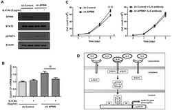
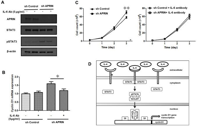
- Figure 4. Treatment with an IL-6-neutralizing Ab P620 attenuates STAT3 activation and cyclin D1 expression, as well as cell proliferation. (A) Western blot analysis. Control (sh control) and APRIN knockdown (sh APRIN) NCI-H460 cells were treated with human IL-6 Ab (5 ug/ml) for 16 h. Protein levels were analyzed by immunoblotting analysis. beta-actin was used as a loading control. (B) Reverse transcription-quantitative PCR assay for cyclin D1 expression. sh control and sh APRIN NCI-H460 cells were treated as in (A) and cyclin D1 mRNA expression. (C) sh control and sh APRIN NCI-H460 cells were treated with P620 human IL-6 Ab (5 ug/ml) every 24 h. Cell number was counted every 24 h. Cell counts without the antibody addition (left) and with the addition of IL-6 Ab (right) are shown. (D) A schematic representation of IL-6/STAT3/cyclin D1 axis in APRIN-associated cellular responses. Increased levels of IL-6 in APRIN-depleted cells binds to its R and activates intracellular STAT3. Activated STAT3 (pSTAT3) dimerizes. The dimers are transported into the nucleus where they enhance the expression of cyclin D1 together with other TFs. *P
- Submitted by
- Invitrogen Antibodies (provider)
- Main image

- Experimental details
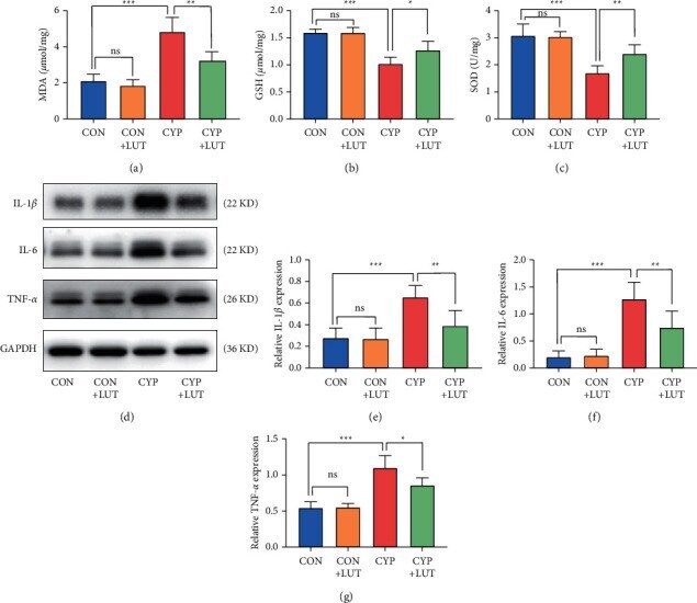
- Figure 2 LUT inhibits the increase in oxidative stress and inflammatory cytokine levels in the bladder tissue caused by CYP. The measurement results of MDA (a), GSH (b), and SOD (c) levels in the bladder of each group. LUT treatment reduced the disorder of bladder oxidative stress-related molecules caused by CYP. (d) The expressions of IL-1 beta , IL-6, and TNF- alpha in the bladder of four groups were detected by western blot. LUT inhibited the upregulation of inflammatory factors induced by CYP. The expression analysis of IL-1 beta (e), IL-6 (f), and TNF- alpha (g) protein in the bladder of four groups ( n = 6). *P < 0.05, ** P < 0.01, and *** P < 0.001; NS: not significant.
- Submitted by
- Invitrogen Antibodies (provider)
- Main image
- Experimental details
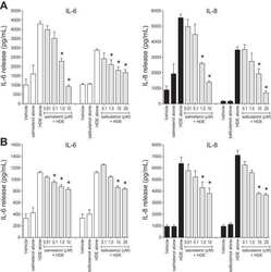
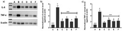
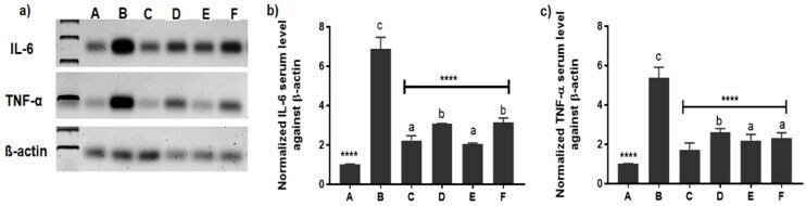
- Figure 8 Western blot demonstration of TNF-alpha and IL-6 expression patterns in the articular cartilages of groups (A-F) 2-week post-treatment. where A is the healthy group, B is the untreated arthritic group, C is the group treated with FLUR-loaded HA-BSA NPs, D is the group treated with blank HA-BSA NPs, E is the group treated with FLUR-loaded BSA NPs and F is the group treated with blank BSA NPs. ( a ) Representative image of western blot showing the expression pattern of IL-6, TNF- alpha and beta-actin, ( b ) Normalized IL-6 expression in all group samples against beta-actin and ( c ) Normalized TNF-alpha expression in all group samples against beta-actin. Statistical significance is shown where **** p
- Submitted by
- Invitrogen Antibodies (provider)
- Main image

- Experimental details
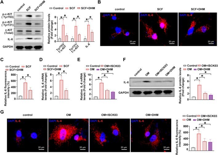
- FIGURE 4 Dihydromyricetin (DHM) represses interleukin-6 (IL-6) through c-KIT inhibition in human valvular interstitial cells (hVICs). (A) IL-6 and phosphorylated c-Kit (sites of Tyr703 and Tyr721) protein levels in hVICs stimulated with stem cell factor (SCF, 100 ng/ml), the c-kit ligand, or cotreated with DHM. One-way ANOVA followed by Bonferroni post hoc test . (B) Immunofluorescent staining was used to detect the protein levels of IL-6 in hVICs following different conditioned culturing conditions. (C) Semiquantification of the fluorescence intensity of IL-6. One-way ANOVA followed by Bonferroni post hoc test. One-way ANOVA followed by Bonferroni post hoc test . (D) mRNA level of IL-6 in hVICs stimulated with SCF and then treated with or without DHM. One-way ANOVA followed by Bonferroni post hoc test . (E) IL-6 mRNA levels in hVICs stimulated with osteogenic induction medium (OM), or cotreated with ISCK03 or DHM. One-way ANOVA followed by Bonferroni post hoc test . (F) IL-6 protein levels in hVICs stimulated with OM or cotreated with ISCK03 or DHM. One-way ANOVA followed by Bonferroni post hoc test . (G) Immunofluorescent staining was used to detect the protein level of IL-6 in hVICs following different conditioned culturing conditions. One-way ANOVA followed by Bonferroni post hoc test . N = 3 per group. Values are the mean +- SD. * p < 0.05 indicates a significant difference.