 Explore
 Validate
 Learn
Learn Western blot
 ELISA
ELISAAntibody data
- Antibody Data
- Antigen structure
- References [1]
- Comments [0]
- Validations
- Western blot [3]
- Other assay [1]
Submit
Validation data
Reference
Comment
Report error
- Product number
- PA1-26811 - Provider product page

- Provider
- Invitrogen Antibodies
- Product name
- IL-6 Polyclonal Antibody
- Antibody type
- Polyclonal
- Antigen
- Recombinant full-length protein
- Description
- This antibody detects recombinant and native IL-6 present in body fluids and cell supernatants in various assays (ie. IL-1 stimulated IL-6 production from fibroblasts). In Western blot analysis of natural cell products or human body fluids, multiple bands of IL-6 will appear due to the variable amount of glycosylation on the molecule.
- Reactivity
- Human
- Host
- Rabbit
- Isotype
- IgG
- Vial size
- 200 µL
- Concentration
- 80 mg/mL
- Storage
- Store at 4°C short term. For long term storage, store at -20°C, avoiding freeze/thaw cycles.
Submitted references Human Mesenchymal Stem Cell-Derived Miniature Joint System for Disease Modeling and Drug Testing.
Li Z, Lin Z, Liu S, Yagi H, Zhang X, Yocum L, Romero-Lopez M, Rhee C, Makarcyzk MJ, Yu I, Li EN, Fritch MR, Gao Q, Goh KB, O'Donnell B, Hao T, Alexander PG, Mahadik B, Fisher JP, Goodman SB, Bunnell BA, Tuan RS, Lin H
Advanced science (Weinheim, Baden-Wurttemberg, Germany) 2022 Jul;9(21):e2105909
Advanced science (Weinheim, Baden-Wurttemberg, Germany) 2022 Jul;9(21):e2105909
No comments: Submit comment
Supportive validation
- Submitted by
- Invitrogen Antibodies (provider)
- Main image
- Experimental details
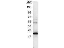
- Western blot detection of IL-6 in 1 µg of recombinant human IL-6. Sample was resolved on a 4-20% Tris-Glycine gel by SDS-PAGE and transferred onto nitrocellulose. After transfer, the membrane was blocked for 30 minutes with 1% BSA-TBST and probed using an IL-6 polyclonal antibody (Product # PA1-26811). Secondary detection was performed using a peroxidase conjugated anti-Rabbit IgG at a 1:40,000 dilution in blocking buffer for 30 min at room temperature, followed by a chemiluminescent substrate. The blot shows detection of a band ~21 kDa in size corresponding to anti-IL6 antibody.

- Submitted by
- Invitrogen Antibodies (provider)
- Main image
- Experimental details
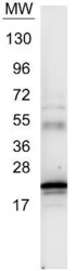
- Western blot using IL-6 Polyclonal Antibody (Product # PA1-26811). 1 µg of our control protein recombinant human IL-6 was resolved on a 4-20% Tris-Glycine gel by SDS-PAGE and transferred onto nitrocellulose. The blot shows detection of a band ~21 kDa in size corresponding to anti-IL6 antibody. Molecular weight markers are also shown (MW). After transfer, the membrane was blocked for 30 minutes with 1% BSA-TBST. Detection occurred using a peroxidase conjµgated anti-Rabbit IgG secondary antibody diluted 1:40,000 in blocking buffer for 30 min at RT followed by reaction with chemiluminescent substrate.

- Submitted by
- Invitrogen Antibodies (provider)
- Main image

- Experimental details
- Western blot was performed using Anti-IL-6 Polyclonal Antibody (Product # PA1-26811) and a 24 kDa band corresponding to IL-6 was observed across to be upregulated upon treatment of HUVEC cells with LPS and Brefeldin A. Whole cell extracts (50 µg lysate) of HUVEC (Lane 1) and HUVEC treated with LPS and Brefeldin A (0.5 µg/mL LPS for 24 h and 300 ng/mL Brefeldin A for last 20 h of LPS stimulation) (Lane 2) were electrophoresed using NuPAGE™ 4-12% Bis-Tris Protein Gel (Product # NP0321BOX). Resolved proteins were then transferred onto a Nitrocellulose membrane (Product # IB23001) by iBlot® 2 Dry Blotting System (Product # IB21001). The blot was probed with the primary antibody (1:1000 dilution) and detected by chemiluminescence with Goat anti-Rabbit IgG (H+L) Superclonal™ Recombinant Secondary Antibody, HRP (Product # A27036, 1:4000 dilution) using the iBright FL 1000 (Product # A32752). Chemiluminescent detection was performed using SuperSignal™ West Dura Extended Duration Substrate (Product # 34076).Expression of IL-6 was observed to be to be upregulated upon treatment of HUVEC cells with LPS and Brefeldin A, HaCaT cells with Poly(I:C) and PTI, and Poly(dA:dT) and PTI.

Supportive validation
- Submitted by
- Invitrogen Antibodies (provider)
- Main image

- Experimental details
- Testing the "therapeutic efficacy" of naproxen (NPX) in the inflamed miniJoint. a) Schematic and timeline of "systemic" administration of NPX in miniJoint (analogous to enteral/parenteral administration in vivo). NPX was added to all the medium streams (indicated by black arrows) after 3 days of IL-1 beta treatment of the SFT tissue. b) Heat map generated from RNA-Seq showing the relative expression of selected marker genes in all four tissues. c) Levels of selected biomarkers in SM, collected from inflamed miniJoint without (IL-1 beta ) or with NPX treatment (IL-1 beta +NPX). The concentration of each marker was normalized to that in the non-NPX-treated IL-1 beta group, with # indicating no statistical difference between the two groups ( p >= 0.05). Data were analyzed by the Student's t -test ( N = 3 biological replicates). The box limits indicate the minimum and maximum values, with the line inside denoting the median. d) Immunostaining images showing reduced levels of MMP-13 and IL-6 in the SFT microtissue after NPX treatment. Scale bar = 50 um. e) Safranin O staining and immunostaining showing more GAG retention and lower MMP-13 level in OC-C, respectively, after NPX treatment. Scale bar = 50 um.
