Explore
 Validate
 Learn
Learn Western blot
 ELISA
ELISAAntibody data
- Antibody Data
- Antigen structure
- References [2]
- Comments [0]
- Validations
- ELISA [1]
- Immunocytochemistry [2]
- Flow cytometry [6]
- Other assay [3]
Submit
Validation data
Reference
Comment
Report error
- Product number
- 701028 - Provider product page

- Provider
- Invitrogen Antibodies
- Product name
- IL-6 Recombinant Rabbit Monoclonal Antibody (4H16L21)
- Antibody type
- Monoclonal
- Antigen
- Other
- Description
- 701028 was successfully to detect IL-6 in Flow application. Intact IgG appears on a non-reducing gel as ~150 kDa band and upon reduction generating a ~25 kDa light chain band and a ~50 kDa heavy chain. Recombinant rabbit monoclonal antibodies are produced using in vitro expression systems. The expression systems are developed by cloning in the specific antibody DNA sequences from immunoreactive rabbits. Then, individual clones are screened to select the best candidates for production. The advantages of using recombinant rabbit monoclonal antibodies include: better specificity and sensitivity, lot-to-lot consistency, animal origin-free formulations, and broader immunoreactivity to diverse targets due to larger rabbit immune repertoire.
- Reactivity
- Human, Mouse
- Host
- Rabbit
- Isotype
- IgG
- Antibody clone number
- 4H16L21
- Vial size
- 100 μg
- Concentration
- 0.5 mg/mL
- Storage
- Store at 4°C short term. For long term storage, store at -20°C, avoiding freeze/thaw cycles.
Submitted references Effect of PM2.5 on invasion and proliferation of HeLa cells and the expression of inflammatory cytokines IL-1 and IL-6.
Four assay designs and on-chip calibration: gadgets for a sepsis protein array.
Huang K, Li W, Chen Y, Zhu J
Oncology letters 2018 Dec;16(6):7068-7073
Oncology letters 2018 Dec;16(6):7068-7073
Four assay designs and on-chip calibration: gadgets for a sepsis protein array.
Buchegger P, Preininger C
Analytical chemistry 2014 Mar 18;86(6):3174-80
Analytical chemistry 2014 Mar 18;86(6):3174-80
No comments: Submit comment
Supportive validation
- Submitted by
- Invitrogen Antibodies (provider)
- Main image

- Experimental details
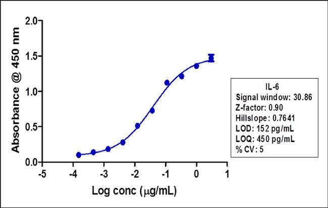
- Indirect ELISA was performed using various dilutions of Anti-IL-6 Rabbit Monoclonal Antibody (Product # 701028) to detect IL-6 recombinant protein coated onto the plate. A non-linear regression analysis was performed (4 PL) and LOD and LOQ for the antibody was determined.
Supportive validation
- Submitted by
- Invitrogen Antibodies (provider)
- Main image

- Experimental details
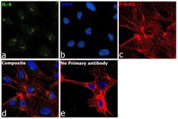
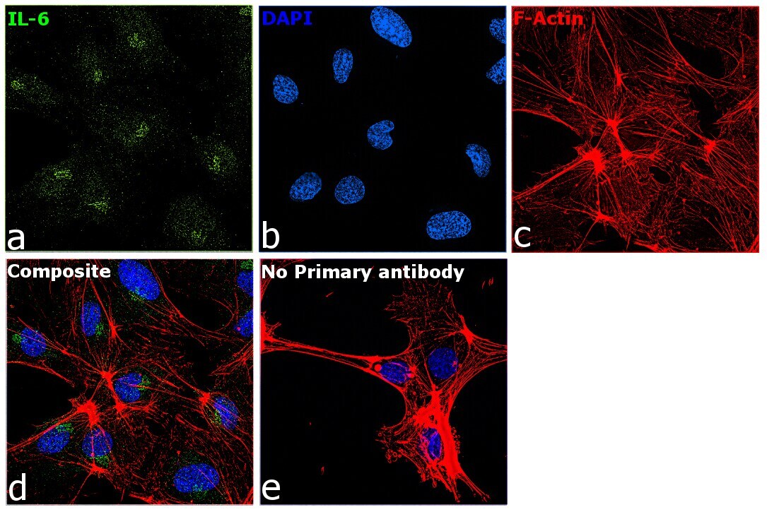
- Immunofluorescence analysis of IL-6 was performed using 70% confluent log phase HUVEC cells. The cells were fixed with 4% paraformaldehyde for 10 minutes, permeabilized with 0.1% Triton™ X-100 for 15 minutes, and blocked with 2% BSA for 45 minutes at room temperature. The cells were labeled with IL-6 Recombinant Rabbit Monoclonal Antibody (4H16L21) (Product # 701028) at 1:200 dilution in 0.1% BSA, incubated at 4 degree celsius overnight and then labeled with Donkey anti-Rabbit IgG (H+L) Highly Cross-Adsorbed Secondary Antibody, Alexa Fluor Plus 488 (Product # A32790), (1:2000 dilution), for 45 minutes at room temperature (Panel a: Green). Nuclei (Panel b:Blue) were stained with ProLong™ Diamond Antifade Mountant with DAPI (Product # P36962). F-actin (Panel c: Red) was stained with Rhodamine Phalloidin (Product # R415, 1:300). Panel d represents the merged image showing predominantly Golgi Complex and Endoplasmic Reticulum localization. Panel e represents control cells with no primary antibody to assess background. The images were captured at 60X magnification.

- Submitted by
- Invitrogen Antibodies (provider)
- Main image
- Experimental details
- Immunofluorescence analysis of IL-6 was performed using 70% confluent log phase HUVEC cells. The cells were fixed with 4% paraformaldehyde for 10 minutes, permeabilized with 0.1% Triton™ X-100 for 15 minutes, and blocked with 2% BSA for 45 minutes at room temperature. The cells were labeled with IL-6 Recombinant Rabbit Monoclonal Antibody (4H16L21) (Product # 701028) at 1:200 dilution in 0.1% BSA, incubated at 4 degree celsius overnight and then labeled with Donkey anti-Rabbit IgG (H+L) Highly Cross-Adsorbed Secondary Antibody, Alexa Fluor Plus 488 (Product # A32790), (1:2000 dilution), for 45 minutes at room temperature (Panel a: Green). Nuclei (Panel b:Blue) were stained with ProLong™ Diamond Antifade Mountant with DAPI (Product # P36962). F-actin (Panel c: Red) was stained with Rhodamine Phalloidin (Product # R415, 1:300). Panel d represents the merged image showing predominantly Golgi Complex and Endoplasmic Reticulum localization. Panel e represents control cells with no primary antibody to assess background. The images were captured at 60X magnification.
Supportive validation
- Submitted by
- Invitrogen Antibodies (provider)
- Main image
- Experimental details
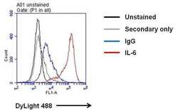
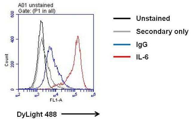
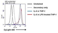
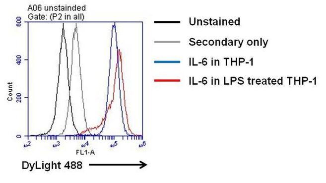
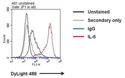
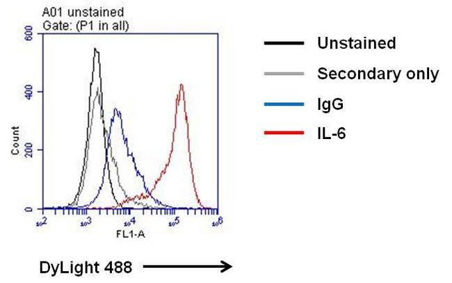
- Flow cytometry analysis of IL-6 on LPS treated THP-1 cells. Cells were fixed with 4% formaldehyde for 30 min on ince and permeabilized with IC permeabilization buffer (Product # PB001). After incubation with blocking buffer (Product # 37525) for 30 min on ice, cells were then stained with anti-IL-6 mouse monoclonal antibody (Product # 701028) or IgG control at 1:100 dilution with IC permeabilization buffer for 30 min on ice. After washing with ice-cold IC permeabilization buffer for 3 times, the cells were stained with DyLight 488 goat anti-mouse secondary antibody (Product # 35502) for 30 min on ice. A representative 10,000 cells were acquired for each sample.
- Submitted by
- Invitrogen Antibodies (provider)
- Main image

- Experimental details
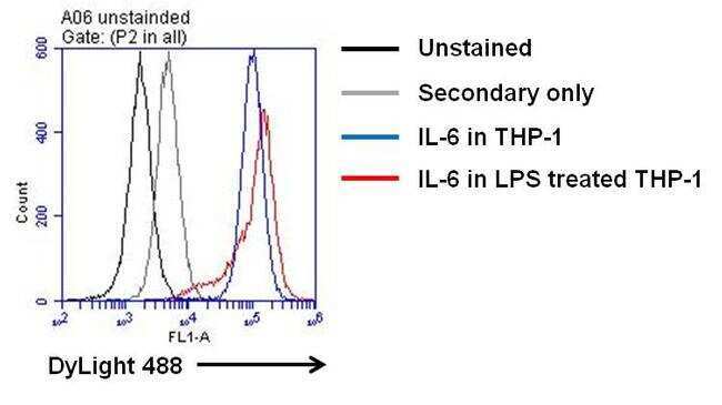
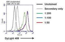
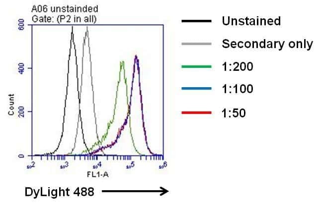
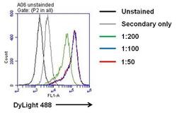
- Flow cytometry analysis of IL-6 on PHA-M stimulated THP-1 cells with or without LPS treatment. Cells were fixed with 4% formaldehyde for 30 min on ince and permeabilized with IC permeabilization buffer (Product # PB001). After incubation with blocking buffer (Product # 37525) for 30 min on ice, cells were then stained with anti-IL-6 rabbit monoclonal antibody (Product # 701028) at indicated dilution with IC permeabilization buffer for 30 min on ice. After washing with ice-cold IC permeabilization buffer for 3 times, the cells were stained with DyLight 488 goat anti-rabbit secondary antibody (Product # 35552) for 30 min on ice. A representative 10,000 cells were acquired for each sample.
- Submitted by
- Invitrogen Antibodies (provider)
- Main image
- Experimental details
- Flow cytometry analysis of IL-6 on LPS treated THP-1 cells. Cells were fixed with 4% formaldehyde for 30 min on ince and permeabilized with IC permeabilization buffer (Product # PB001). After incubation with blocking buffer (Product # 37525) for 30 min on ice, cells were then stained with anti-IL-6 rabbit monoclonal antibody (Product # 701028) at indicated dilution with IC permeabilization buffer for 30 min on ice. After washing with ice-cold IC permeabilization buffer for 3 times, the cells were stained with DyLight 488 goat anti-rabbit secondary antibody (Product # 35552) for 30 min on ice. A representative 10,000 cells were acquired for each sample.
- Submitted by
- Invitrogen Antibodies (provider)
- Main image
- Experimental details
- Flow cytometry analysis of IL-6 on PHA-M stimulated THP-1 cells with or without LPS treatment. Cells were fixed with 4% formaldehyde for 30 min on ince and permeabilized with IC permeabilization buffer (Product # PB001). After incubation with blocking buffer (Product # 37525) for 30 min on ice, cells were then stained with anti-IL-6 rabbit monoclonal antibody (Product # 701028) at indicated dilution with IC permeabilization buffer for 30 min on ice. After washing with ice-cold IC permeabilization buffer for 3 times, the cells were stained with DyLight 488 goat anti-rabbit secondary antibody (Product # 35552) for 30 min on ice. A representative 10,000 cells were acquired for each sample.
- Submitted by
- Invitrogen Antibodies (provider)
- Main image
- Experimental details
- Flow cytometry analysis of IL-6 on LPS treated THP-1 cells. Cells were fixed with 4% formaldehyde for 30 min on ince and permeabilized with IC permeabilization buffer (Product # PB001). After incubation with blocking buffer (Product # 37525) for 30 min on ice, cells were then stained with anti-IL-6 rabbit monoclonal antibody (Product # 701028) at indicated dilution with IC permeabilization buffer for 30 min on ice. After washing with ice-cold IC permeabilization buffer for 3 times, the cells were stained with DyLight 488 goat anti-rabbit secondary antibody (Product # 35552) for 30 min on ice. A representative 10,000 cells were acquired for each sample.

- Submitted by
- Invitrogen Antibodies (provider)
- Main image
- Experimental details
- Flow cytometry analysis of IL-6 on LPS treated THP-1 cells. Cells were fixed with 4% formaldehyde for 30 min on ince and permeabilized with IC permeabilization buffer (Product # PB001). After incubation with blocking buffer (Product # 37525) for 30 min on ice, cells were then stained with anti-IL-6 mouse monoclonal antibody (Product # 701028) or IgG control at 1:100 dilution with IC permeabilization buffer for 30 min on ice. After washing with ice-cold IC permeabilization buffer for 3 times, the cells were stained with DyLight 488 goat anti-mouse secondary antibody (Product # 35502) for 30 min on ice. A representative 10,000 cells were acquired for each sample.
Supportive validation
- Submitted by
- Invitrogen Antibodies (provider)
- Main image

- Experimental details
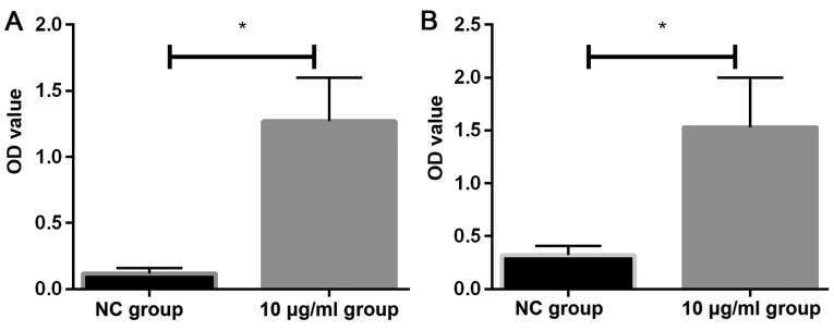
- Figure 5. ELISA. ELISA was used to detect the expression of IL-1 and IL-6 in two groups of cells. The OD values of IL-1 and IL-6 in HeLa cells treated with 10 ug/ml PM2.5 were significantly higher than those in NC group. *P
- Submitted by
- Invitrogen Antibodies (provider)
- Main image

- Experimental details
- Indirect ELISA was performed using various dilutions of Anti-IL-6 Rabbit Monoclonal Antibody (Product # 701028) to detect IL-6 recombinant protein coated onto the plate. A non-linear regression analysis was performed (4 PL) and LOD and LOQ for the antibody was determined.

- Submitted by
- Invitrogen Antibodies (provider)
- Main image
- Experimental details
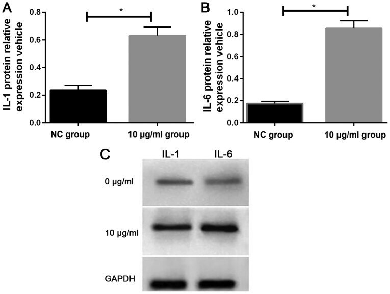
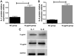
- Figure 3. Western blotting. Compared with HeLa cells in NC group, the relative expression levels of (A) IL-1 and (B) IL-6 proteins in HeLa cells treated with 10 ug/ml PM2.5 were significantly increased. (C) Western blots of IL-1 and IL-6 protein in the two groups of cells are shown. *P