Explore
 Validate
 Learn
Learn Western blot
 ELISA
ELISAAntibody data
- Antibody Data
- Antigen structure
- References [0]
- Comments [0]
- Validations
- Western blot [1]
- Immunohistochemistry [3]
Submit
Validation data
Reference
Comment
Report error
- Product number
- LS-C154128 - Provider product page

- Provider
- LSBio
- Product name
- IL6 / Interleukin 6 Antibody LS-C154128
- Antibody type
- Polyclonal
- Description
- Delipidation, salt fractionation and ion exchange chromatography followed by dialysis.
- Reactivity
- Mouse
- Host
- Rabbit
- Isotype
- IgG
- Storage
- Store lyophilized at 4°C. Once reconstituted, aliquot and store at -20°C. Avoid freeze/thaw cycles. Store undiluted.
No comments: Submit comment
Enhanced validation
- Submitted by
- LSBio (provider)
- Enhanced method
- Genetic validation
- Main image
- Experimental details
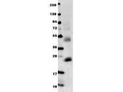
- Anti-Mouse IL-6 Antibody - Western Blot. Anti-mouse IL-6 antibody in western blot shows detection of recombinant mouse IL-6 raised in E. coli. Recombinant truncated protein (0.1 ug, 21.7 kD) was loaded on to an SDS-PAGE gel, and after separation, transferred to nitrocellulose. The membrane was blocked with 1% BSA in TBST for 30 min at RT, followed by incubation with Anti-Mouse IL-6 antibody diluted 1:1000 in 1% BSA in TBST overnight at 4°C. After washes, the blot was reacted with secondary antibody Dylight 649 Conjugated Anti-Rabbit IgG (H&L) (Goat) Antibody ( diluted 1:20000 in blocking buffer (MB-070) for 30 min. at RT. Data was collected using Bio-Rad VersaDoc 4000 MP imaging system.

Enhanced validation
- Submitted by
- LSBio (provider)
- Enhanced method
- Genetic validation
- Main image
- Experimental details
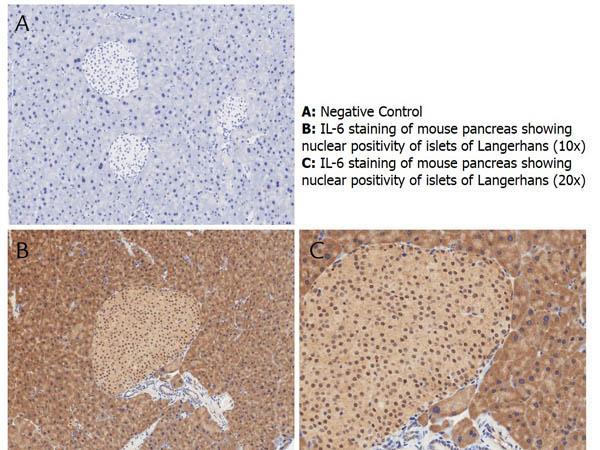
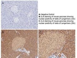
- Immunohistochemistry with anti-IL-6 antibody showing nuclear positivity of islets of Langerhans (brown staining) and cytoplasmic staining in mouse pancreas at 10x and 20x (B & C). Staining was performed on Leica Bond system using the standard protocol. Formalin fixed/paraffin embedded tissue sections were subjected to antigen retrieval with E1 (Leica Microsystems) retrieval solution for 20 min and then incubated with rabbit anti-mouse IL-6 antibody at 1:50 dilution for 60 minutes. Biotinylated Anti-rabbit secondary antibody was used at 1:200 dilution to detect primary antibody. The reaction was developed using streptavidin-HRP conjugated compact polymer system and visualized with chromogen substrate, 3’3-diamino-benzidine substrate (DAB). The sections were then counterstained with hematoxylin to detect cell nuclei.

- Submitted by
- LSBio (provider)
- Enhanced method
- Genetic validation
- Main image

- Experimental details
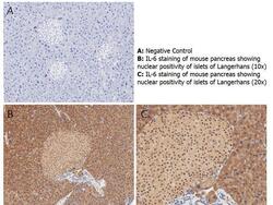
- Immunohistochemistry with anti-IL-6 antibody showing nuclear positivity of islets of Langerhans (brown staining) and cytoplasmic staining in mouse pancreas at 10x and 20x (B & C). Staining was performed on Leica Bond system using the standard protocol. Formalin fixed/paraffin embedded tissue sections were subjected to antigen retrieval with E1 (Leica Microsystems) retrieval solution for 20 min and then incubated with rabbit anti-mouse IL-6 antibody at 1:50 dilution for 60 minutes. Biotinylated Anti-rabbit secondary antibody was used at 1:200 dilution to detect primary antibody. The reaction was developed using streptavidin-HRP conjugated compact polymer system and visualized with chromogen substrate, 3’3-diamino-benzidine substrate (DAB). The sections were then counterstained with hematoxylin to detect cell nuclei.
- Submitted by
- LSBio (provider)
- Main image
- Experimental details
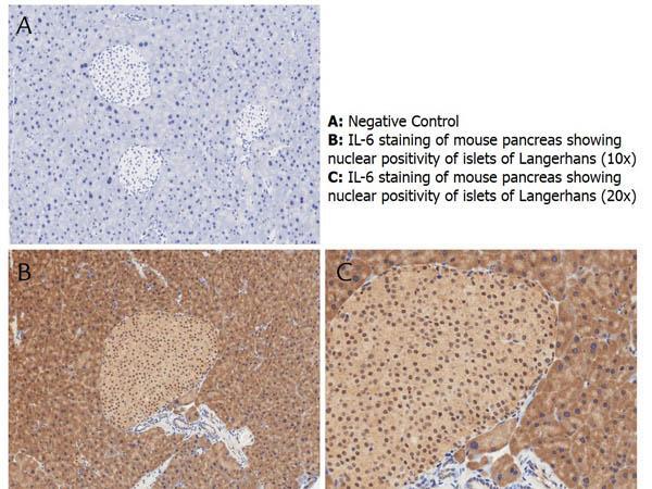
- Immunohistochemistry with anti-IL-6 antibody showing nuclear positivity of islets of Langerhans (brown staining) and cytoplasmic staining in mouse pancreas at 10x and 20x (B & C). Staining was performed on Leica Bond system using the standard protocol. Formalin fixed/paraffin embedded tissue sections were subjected to antigen retrieval with E1 (Leica Microsystems) retrieval solution for 20 min and then incubated with rabbit anti-mouse IL-6 antibody at 1:50 dilution for 60 minutes. Biotinylated Anti-rabbit secondary antibody was used at 1:200 dilution to detect primary antibody. The reaction was developed using streptavidin-HRP conjugated compact polymer system and visualized with chromogen substrate, 3’3-diamino-benzidine substrate (DAB). The sections were then counterstained with hematoxylin to detect cell nuclei.