 Explore
 Validate
 Learn
Learn Western blot
Antibody data
- Antibody Data
- Antigen structure
- References [0]
- Comments [0]
- Validations
- Western blot [2]
- Immunocytochemistry [1]
- Immunohistochemistry [1]
Submit
Validation data
Reference
Comment
Report error
- Product number
- ABIN2508605 - Provider product page

- Provider
- antibodies-online
- Product name
- anti-Interleukin 6 (IL6) antibody (Biotin)
- Antibody type
- Polyclonal
- Antigen
- Other
- Description
- Produced from sera of goats pre-immunized with highly pure recombinant Human IL-6. Anti-Human IL-6 specific antibody was purified by affinity chromatography and then biotinylated.
- Reactivity
- Human
- Host
- Goat
- Conjugate
- Biotin
- Vial size
- 50 μg
- Storage
- -20°C
No comments: Submit comment
Supportive validation
- Submitted by
- antibodies-online (provider)
- Main image

- Experimental details
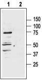
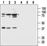
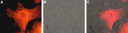
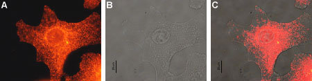
- Western blot analysis of human normal skin fibroblast cell line Malme-3 (lanes 1 and 4) and human malignant melanoma cell lines Malme-3M (lanes 2 and 5) and A875 (lanes 3 and 6): 1, 2, 3. Anti-Melanocortin Receptor 1 antibody (ABIN2511173), (1:500). 4, 5, 6. Anti-Melanocortin Receptor 1 antibody, preincubated with the control peptide antigen Western blot analysis of rat adrenal lysate: 1. Anti-Melanocortin Receptor 1 antibody (ABIN2511173), (1:400). 2. Anti-Melanocortin Receptor 1 antibody, preincubated with the control peptide antigen. Expression of MC1R in normal skin and melanoma Immunohistochemical staining of paraffin embedded normal skin and melanoma sections using Anti-Melanocortin Receptor 1 antibody (ABIN2511173) (1:100). MCR1 staining (red-brown color is highly specific in A. epidermal cells, B. eccrine sweat gland cells and C. melanoma cells. Color reaction was obtained with DAB. Hematoxilin is used as the counterstain. Expression of MC1R in human Malme-3M cells Immunocytochemical staining of human paraformaldehyde fixed and permeabilized malignant melanoma cell lines (Malme-3M). A. Cells were stained with Anti-Melanocortin Receptor 1 antibody (ABIN2511173), (1:200) followed by goat-anti-rabbit- AlexaFluor-555 secondary antibody. B. Live view of the same field as in (A). C. Computer merge of (A) and (B).
- Submitted by
- antibodies-online (provider)
- Main image

- Experimental details
- Western blot analysis of human normal skin fibroblast cell line Malme-3 (lanes 1 and 4) and human malignant melanoma cell lines Malme-3M (lanes 2 and 5) and A875 (lanes 3 and 6): 1, 2, 3. Anti-Melanocortin Receptor 1 antibody (ABIN2511173), (1:500). 4, 5, 6. Anti-Melanocortin Receptor 1 antibody, preincubated with the control peptide antigen Western blot analysis of rat adrenal lysate: 1. Anti-Melanocortin Receptor 1 antibody (ABIN2511173), (1:400). 2. Anti-Melanocortin Receptor 1 antibody, preincubated with the control peptide antigen. Expression of MC1R in normal skin and melanoma Immunohistochemical staining of paraffin embedded normal skin and melanoma sections using Anti-Melanocortin Receptor 1 antibody (ABIN2511173) (1:100). MCR1 staining (red-brown color is highly specific in A. epidermal cells, B. eccrine sweat gland cells and C. melanoma cells. Color reaction was obtained with DAB. Hematoxilin is used as the counterstain. Expression of MC1R in human Malme-3M cells Immunocytochemical staining of human paraformaldehyde fixed and permeabilized malignant melanoma cell lines (Malme-3M). A. Cells were stained with Anti-Melanocortin Receptor 1 antibody (ABIN2511173), (1:200) followed by goat-anti-rabbit- AlexaFluor-555 secondary antibody. B. Live view of the same field as in (A). C. Computer merge of (A) and (B).
Supportive validation
- Submitted by
- antibodies-online (provider)
- Main image
- Experimental details
- Image(s): Immunofluorescence
Supportive validation
- Submitted by
- antibodies-online (provider)
- Main image

- Experimental details
- Image(s): Immunohistochemistry
