Explore
 Validate
 Learn
Learn Western blot
Antibody data
- Antibody Data
- Antigen structure
- References [4]
- Comments [0]
- Validations
- Western blot [2]
- Immunohistochemistry [1]
Submit
Validation data
Reference
Comment
Report error
- Product number
- AP7932a - Provider product page

- Provider
- Abcepta
- Proper citation
- Abgent Cat#AP7932a, RRID:AB_2163805
- Product name
- PIM1 Antibody (C-term)
- Antibody type
- Polyclonal
- Antigen
- Synthetic peptide
- Description
- Purified Rabbit Polyclonal Antibody (Pab)
- Reactivity
- Human
- Host
- Rabbit
- Isotype
- IgG
- Vial size
- 400 µl
- Storage
- Maintain refrigerated at 2-8°C for up to 6 months. For long term storage store at -20°C in small aliquots to prevent freeze-thaw cycles.
Submitted references Prognostic impact of protein overexpression of the proto-oncogene PIM-1 in gastric cancer.
Hypoxia-mediated up-regulation of Pim-1 contributes to solid tumor formation.
Hypoxia-inducible proto-oncogene Pim-1 is a prognostic marker in pancreatic ductal adenocarcinoma.
Comprehensive identification of proteins in Hodgkin lymphoma-derived Reed-Sternberg cells by LC-MS/MS.
Warnecke-Eberz U, Bollschweiler E, Drebber U, Metzger R, Baldus SE, Hölscher AH, Mönig S
Anticancer research 2009 Nov;29(11):4451-5
Anticancer research 2009 Nov;29(11):4451-5
Hypoxia-mediated up-regulation of Pim-1 contributes to solid tumor formation.
Chen J, Kobayashi M, Darmanin S, Qiao Y, Gully C, Zhao R, Kondo S, Wang H, Wang H, Yeung SC, Lee MH
The American journal of pathology 2009 Jul;175(1):400-11
The American journal of pathology 2009 Jul;175(1):400-11
Hypoxia-inducible proto-oncogene Pim-1 is a prognostic marker in pancreatic ductal adenocarcinoma.
Reiser-Erkan C, Erkan M, Pan Z, Bekasi S, Giese NA, Streit S, Michalski CW, Friess H, Kleeff J
Cancer biology & therapy 2008 Sep;7(9):1352-9
Cancer biology & therapy 2008 Sep;7(9):1352-9
Comprehensive identification of proteins in Hodgkin lymphoma-derived Reed-Sternberg cells by LC-MS/MS.
Wallentine JC, Kim KK, Seiler CE 3rd, Vaughn CP, Crockett DK, Tripp SR, Elenitoba-Johnson KS, Lim MS
Laboratory investigation; a journal of technical methods and pathology 2007 Nov;87(11):1113-24
Laboratory investigation; a journal of technical methods and pathology 2007 Nov;87(11):1113-24
No comments: Submit comment
Supportive validation
- Submitted by
- Abcepta (provider)
- Main image
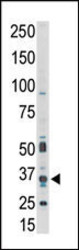
- Experimental details
- Western blot analysis of anti-PIM1 Pab (Cat. #AP7932a) in A549 cell lysate. PIM1 (arrow) was detected using purified Pab. Secondary HRP-anti-rabbit was used for signal visualization with chemiluminescence.
- Primary Ab dilution
- 1:1000

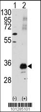
- Submitted by
- Abcepta (provider)
- Main image

- Experimental details
- Western blot analysis of PIM1 (arrow) using PIM1 Antibody (C-term) (Cat.#AP7932a). 293 cell lysates (2 ug/lane) either nontransfected (Lane 1) or transiently transfected with the PIM1 gene (Lane 2) (Origene Technologies).
- Primary Ab dilution
- 1:1000
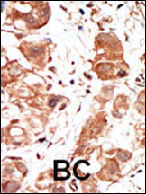
Supportive validation
- Submitted by
- Abcepta (provider)
- Main image

- Experimental details
- "Formalin-fixed and paraffin-embedded human cancer tissue reacted with the primary antibody, which was peroxidase-conjugated to the secondary antibody, followed by DAB staining. This data demonstrates the use of this antibody for immunohistochemistry; clinical relevance has not been evaluated. BC = breast carcinoma; HC = hepatocarcinoma."
- Primary Ab dilution
- 1:50~100