Explore
 Validate
 Learn
Learn Western blot
 ELISA
ELISA Chromatin Immunoprecipitation
Antibody data
- Antibody Data
- Antigen structure
- References [5]
- Comments [0]
- Validations
- Chromatin Immunoprecipitation [2]
- Other assay [6]
Submit
Validation data
Reference
Comment
Report error
- Product number
- 39-4600 - Provider product page

- Provider
- Invitrogen Antibodies
- Product name
- PIM1 Monoclonal Antibody (ZP003)
- Antibody type
- Monoclonal
- Antigen
- Synthetic peptide
- Description
- 42-4800 has been successfully used in ChIP analysis of PIM1 in human J9.2 J-lat T lymphocytes.
- Reactivity
- Human
- Host
- Mouse
- Isotype
- IgG
- Antibody clone number
- ZP003
- Vial size
- 100 μg
- Concentration
- 0.5 mg/mL
- Storage
- -20°C
Submitted references Pim1 maintains telomere length in mouse cardiomyocytes by inhibiting TGFβ signalling.
PIM1 Promotes Survival of Cardiomyocytes by Upregulating c-Kit Protein Expression.
Functional Effect of Pim1 Depends upon Intracellular Localization in Human Cardiac Progenitor Cells.
Nucleostemin rejuvenates cardiac progenitor cells and antagonizes myocardial aging.
Fibronectin is essential for reparative cardiac progenitor cell response after myocardial infarction.
Ebeid DE, Khalafalla FG, Broughton KM, Monsanto MM, Esquer CY, Sacchi V, Hariharan N, Korski KI, Moshref M, Emathinger J, Cottage CT, Quijada PJ, Nguyen JH, Alvarez R, Völkers M, Konstandin MH, Wang BJ, Firouzi F, Navarrete JM, Gude NA, Goumans MJ, Sussman MA
Cardiovascular research 2021 Jan 1;117(1):201-211
Cardiovascular research 2021 Jan 1;117(1):201-211
PIM1 Promotes Survival of Cardiomyocytes by Upregulating c-Kit Protein Expression.
Ebeid DE, Firouzi F, Esquer CY, Navarrete JM, Wang BJ, Gude NA, Sussman MA
Cells 2020 Aug 31;9(9)
Cells 2020 Aug 31;9(9)
Functional Effect of Pim1 Depends upon Intracellular Localization in Human Cardiac Progenitor Cells.
Samse K, Emathinger J, Hariharan N, Quijada P, Ilves K, Völkers M, Ormachea L, De La Torre A, Orogo AM, Alvarez R, Din S, Mohsin S, Monsanto M, Fischer KM, Dembitsky WP, Gustafsson ÅB, Sussman MA
The Journal of biological chemistry 2015 May 29;290(22):13935-47
The Journal of biological chemistry 2015 May 29;290(22):13935-47
Nucleostemin rejuvenates cardiac progenitor cells and antagonizes myocardial aging.
Hariharan N, Quijada P, Mohsin S, Joyo A, Samse K, Monsanto M, De La Torre A, Avitabile D, Ormachea L, McGregor MJ, Tsai EJ, Sussman MA
Journal of the American College of Cardiology 2015 Jan 20;65(2):133-47
Journal of the American College of Cardiology 2015 Jan 20;65(2):133-47
Fibronectin is essential for reparative cardiac progenitor cell response after myocardial infarction.
Konstandin MH, Toko H, Gastelum GM, Quijada P, De La Torre A, Quintana M, Collins B, Din S, Avitabile D, Völkers M, Gude N, Fässler R, Sussman MA
Circulation research 2013 Jul 5;113(2):115-25
Circulation research 2013 Jul 5;113(2):115-25
No comments: Submit comment
Supportive validation
- Submitted by
- Invitrogen Antibodies (provider)
- Main image
- Experimental details
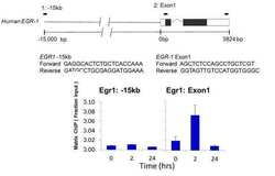
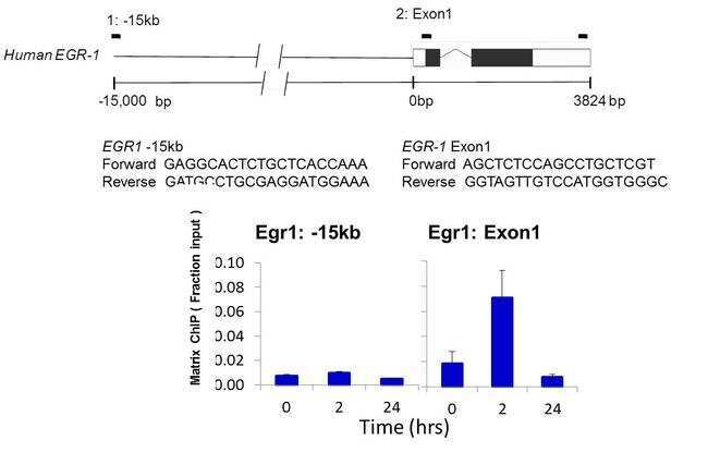
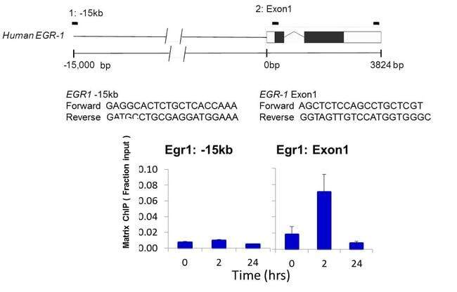
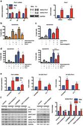
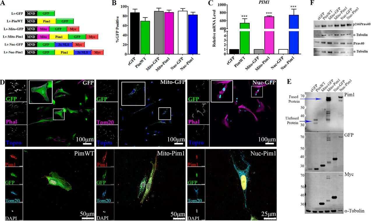
- Chromatin immunoprecipitation analysis of PIM-1 was performed using cross-linked chromatin from Human J9.2 J-lat T lymphocytes culture treated with 10 µg/mL PHA (phytohemaglutin) for 0, 2, and 24 hours. Immunoprecipitation was performed using a multiplex microplate Matrix ChIP assay (see reference for Matrix ChIP protocol: http://www.ncbi.nlm.nih.gov/pubmed/22098709) with 1.0 µL/100 µL well volume of a PIM-1 monoclonal antibody (Product # 39-4600). Chromatin aliquots from ~1 x 10⁴ cells were used per ChIP pull-down. Quantitative PCR data were done in quadruplicate using 1 µL of eluted DNA in 2 µL SYBR real-time PCR reactions containing primers shown to amplify -15kb and exon-1 of the EGR1 gene. PCR calibration curves were generated for each primer pair from a dilution series of sheared total genomic DNA. Quantitation of immunoprecipitated chromatin is presented as signal relative to the total amount of input chromatin. Results represent the mean +/- SEM for three experiments. Schematic representations of the EGR-1 locus are shown above the data where boxes represent exons (black boxes = translated regions, white boxes = untranslated regions), the zigzag line represents an intron, and the straight line represents upstream sequence. Regions amplified by the primers are represented by black bars. Data courtesy of Dr. Karol Bomsztyk at the University of Washington, Seattle, WA.
- Submitted by
- Invitrogen Antibodies (provider)
- Main image

- Experimental details
- Chromatin immunoprecipitation analysis of PIM-1 was performed using cross-linked chromatin from Human J9.2 J-lat T lymphocytes culture treated with 10 µg/mL PHA (phytohemaglutin) for 0, 2, and 24 hours. Immunoprecipitation was performed using a multiplex microplate Matrix ChIP assay (see reference for Matrix ChIP protocol: http://www.ncbi.nlm.nih.gov/pubmed/22098709) with 1.0 µL/100 µL well volume of a PIM-1 monoclonal antibody (Product # 39-4600). Chromatin aliquots from ~1 x 10⁴ cells were used per ChIP pull-down. Quantitative PCR data were done in quadruplicate using 1 µL of eluted DNA in 2 µL SYBR real-time PCR reactions containing primers shown to amplify -15kb and exon-1 of the EGR1 gene. PCR calibration curves were generated for each primer pair from a dilution series of sheared total genomic DNA. Quantitation of immunoprecipitated chromatin is presented as signal relative to the total amount of input chromatin. Results represent the mean +/- SEM for three experiments. Schematic representations of the EGR-1 locus are shown above the data where boxes represent exons (black boxes = translated regions, white boxes = untranslated regions), the zigzag line represents an intron, and the straight line represents upstream sequence. Regions amplified by the primers are represented by black bars. Data courtesy of Dr. Karol Bomsztyk at the University of Washington, Seattle, WA.
Supportive validation
- Submitted by
- Invitrogen Antibodies (provider)
- Main image
- Experimental details
- NULL
- Submitted by
- Invitrogen Antibodies (provider)
- Main image
- Experimental details
- NULL

- Submitted by
- Invitrogen Antibodies (provider)
- Main image
- Experimental details
- NULL
- Submitted by
- Invitrogen Antibodies (provider)
- Main image
- Experimental details
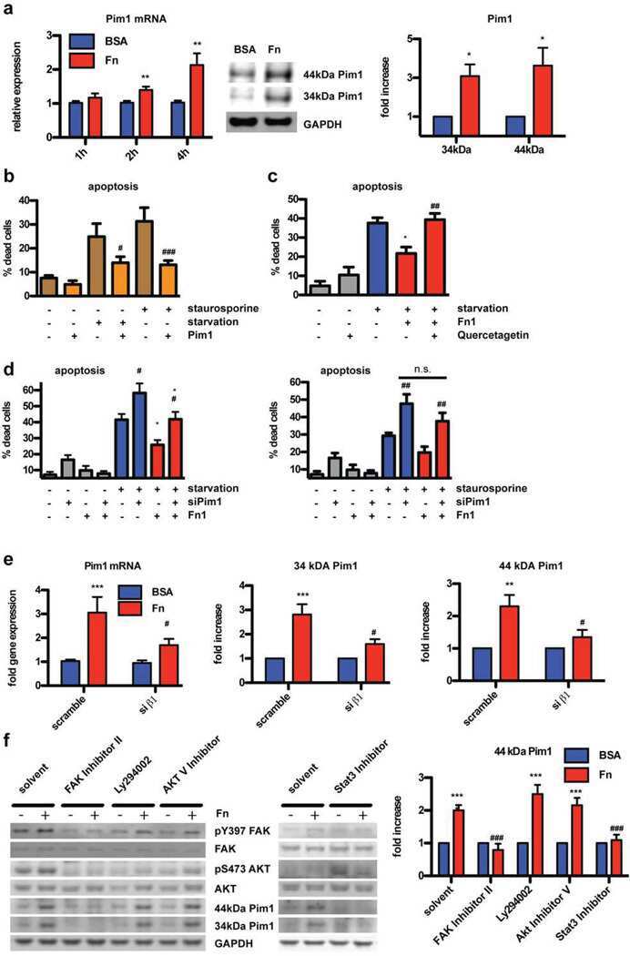
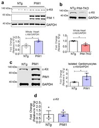
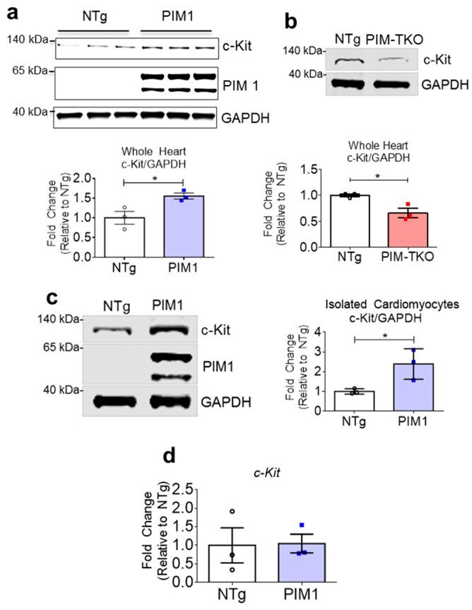
- Figure 1 PIM1 upregulates c-Kit protein expression. ( a ) Immunoblot analysis showing expression of c-Kit in PIM1 vs. NTg whole heart lysates with quantification below. N = 3, Error bars represent SEM, * p < 0.05 vs. untreated as measured by Student t test. ( b ) Immunoblot analysis showing the expression of c-Kit in PIM-TKO vs. NTg whole heart lysates with quantification shown below. N = 3, error bars represent SEM, * p < 0.05 vs. untreated as measured by Student t test. ( c ) Immunoblot analysis showing c-Kit expression in cardiomyocytes isolated from PIM1-overexpressing hearts vs. NTg with quantification on the right. N = 3, error bars represent SEM, * p < 0.05 vs. untreated as measured by Student t test. ( d ) Gene expression of c-Kit in cardiomyocytes isolated from PIM1 and NTg hearts as revealed by qPCR. N = 3, Error bars represent SEM.
- Submitted by
- Invitrogen Antibodies (provider)
- Main image
- Experimental details
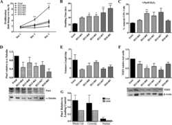
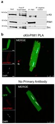
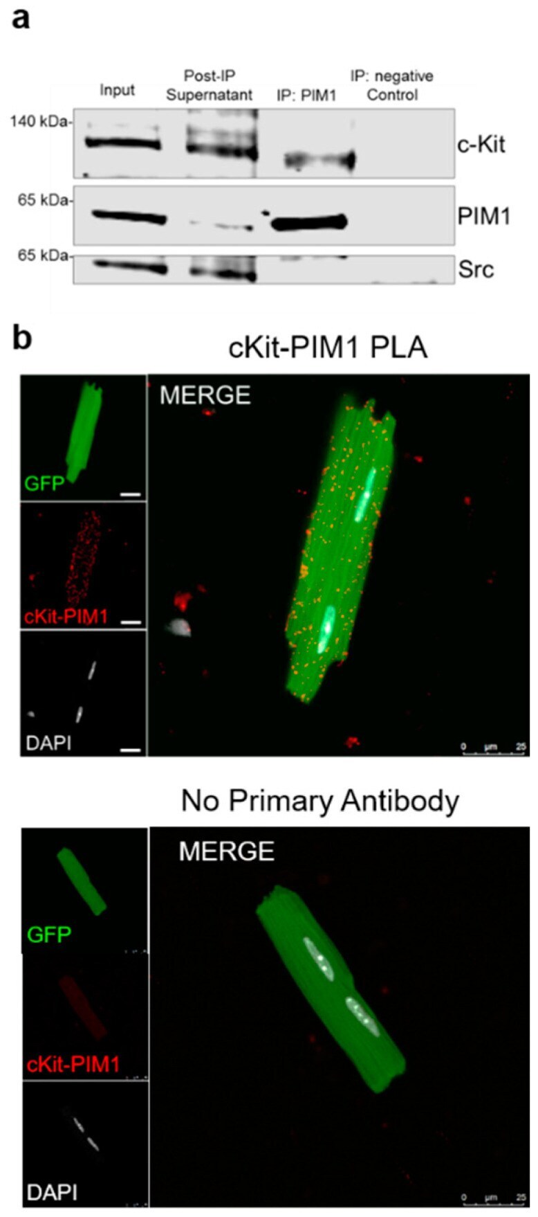
- Figure 2 PIM1 interacts with c-Kit. ( a ) Immunoblot analysis of a co-immunoprecipitation using whole heart lysates from PIM1 hearts in which PIM1 was pulled down. ( b ) Interaction between PIM1 and c-Kit as determined by proximity ligation assay (top panel) and in the absence of primary antibody (bottom panel). Proximity events are shown as red dots.
- Submitted by
- Invitrogen Antibodies (provider)
- Main image
- Experimental details
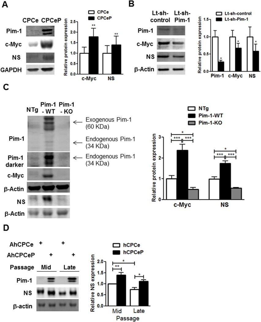
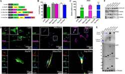
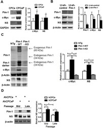
- Pim-1 Regulates c-Myc and NS Pim-1 is necessary and sufficient for c-Myc and NS regulation. Immunoblots and densitometric analyses of Pim-1, c-Myc, and NS in (A) YCPCs expressing express green fluorescent protein (eGFP) (CPCe) or eGFP and Pim-1 (CPCeP; N = 7) (B) YCPCs with knockdown of Pim-1 (Lt-sh-Pim-1, N = 4) (C) Protein lysates from hearts of 3-month-old transgenic mice with cardiac specific overexpression of Pim-1 (Pim-1-WT), global Pim-1 knockout mice (Pim-1-KO), and nontransgenic (NTg) mice (N = 4 each). Exogenous Pim-1 fused to GFP as well as endogenous Pim-1 levels are indicated at 2 different exposure levels. (D) Immunoblots and denistometric analyses of NS in AhCPCs (Line H10-001) with overexpression of Pim-1 (AhCPCeP) or eGFP (AhCPCe) at mid (p5-6) and late (p10) passages (N = 4). *p < 0.05; **p < 0.01; ***p < 0.001 Other abbreviations as in Figures 1 and 2.