Explore
 Validate
 Learn
Learn Western blot
 Immunocytochemistry
Antibody data
- Antibody Data
- Antigen structure
- References [1]
- Comments [0]
- Validations
- Immunocytochemistry [5]
- Flow cytometry [2]
Submit
Validation data
Reference
Comment
Report error
- Product number
- PA1-114 - Provider product page

- Provider
- Invitrogen Antibodies
- Product name
- PRDM14 Polyclonal Antibody
- Antibody type
- Polyclonal
- Antigen
- Recombinant full-length protein
- Reactivity
- Human
- Host
- Rabbit
- Isotype
- IgG
- Vial size
- 100 μg
- Concentration
- 1 mg/mL
- Storage
- -20°C, Avoid Freeze/Thaw Cycles
Submitted references BMP4 is insufficient to differentiate umbilical cord mesenchymal stem cells into germ cell-like cells in vitro.
Wang P, Hu H, Li X, Zhang R, Cheng H, Qin H, Ji G, Feng H, Liu Y, Lin J
Ginekologia polska 2023;94(1):64-72
Ginekologia polska 2023;94(1):64-72
No comments: Submit comment
Supportive validation
- Submitted by
- Invitrogen Antibodies (provider)
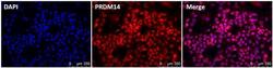
- Main image
- Experimental details
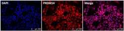
- Immunofluorescence analysis of PRDM14 (red, middle panel) in human embryonic stem cell H9 line grown on irradiated MEF-feeder layer. The cells were fixed with 4% paraformaldehyde at room temperature for 10 min and permeabilized with 0.25% Triton-X 100 for 5 min and blocked with the 10% BSA in PBS for 30 min at 37°C. Cells were stained with a PRDM14 polyclonal antibody (Product # PA1-114) at a dilution of 1:200 in 3% BSA/PBS blocking buffer overnight at 4°C, and then incubated with a RRX-conjugated donkey anti-rabbit IgG secondary antibody at a dilution of 1:500 for 1 hour at room temperature. Nucleus DNA (blue, left panel) was stained with DAPI (Product # D1306). Data courtesy of the innovators program.

- Submitted by
- Invitrogen Antibodies (provider)
- Main image
- Experimental details
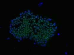
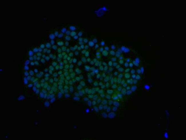
- Immunofluorescence analysis of PRDM14 (green) in a human iPSC line, Gibco Episomal iPSC line (Product # A18945) grown on vitronectin in Essential 8 medium (Product # A1517001). The cells were fixed and permeabilized using Image-IT® Fixation/Permeabilization kit (Product # R37602), and blocked with the including blocking buffer for one hour at room temperature. Cells were stained with a PRDM14 polyclonal antibody (Product # PA1-114) at a dilution of 1:500 in 3% BSA/PBS blocking buffer overnight at 4°C, and then incubated with a DyLight 488-conjugated goat anti-rabbit IgG secondary antibody (Product # 35553) at a dilution of 1:250 for 1 hour at room temperature. Nucleus DNA (blue) was stained with DAPI (Product # D1306). Images were taken on an EVOS® FLoid® Cell Imaging Station at 10X magnification.
- Submitted by
- Invitrogen Antibodies (provider)
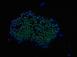
- Main image

- Experimental details
- Immunofluorescence analysis of PRDM14 (green) in a human iPSC line, Gibco Episomal iPSC line (Product # A18945) grown on vitronectin in Essential 8™ medium (Product # A1517001). The cells were fixed and permeabilized using Image-IT® Fixation/Permeabilization kit (Product # R37602), and blocked with the including blocking buffer for one hour at room temperature. Cells were stained with a PRDM14 polyclonal antibody (Product # PA1-114) at a dilution of 1:500 in 3% BSA/PBS blocking buffer overnight at 4°C, and then incubated with a DyLight 488-conjugated goat anti-rabbit IgG secondary antibody (Product # 35553) at a dilution of 1:250 for 1 hour at room temperature. Nucleus DNA (blue) was stained with DAPI (Product # D1306). Images were taken on an EVOS® FLoid® Cell Imaging Station at 10X magnification.

- Submitted by
- Invitrogen Antibodies (provider)
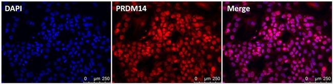
- Main image
- Experimental details
- Immunofluorescence analysis of PRDM14 (red, middle panel) in human embryonic stem cell H9 line grown on irradiated MEF-feeder layer. The cells were fixed with 4% paraformaldehyde at room temperature for 10 min and permeabilized with 0.25% Triton-X 100 for 5 min and blocked with the 10% BSA in PBS for 30 min at 37°C. Cells were stained with a PRDM14 polyclonal antibody (Product # PA1-114) at a dilution of 1:200 in 3% BSA/PBS blocking buffer overnight at 4°C, and then incubated with a RRX-conjugated donkey anti-rabbit IgG secondary antibody at a dilution of 1:500 for 1 hour at room temperature. Nucleus DNA (blue, left panel) was stained with DAPI (Product # D1306). Data courtesy of the innovators program.
- Submitted by
- Invitrogen Antibodies (provider)
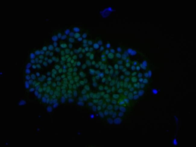
- Main image
- Experimental details
- Immunofluorescence analysis of PRDM14 (green) in a human iPSC line, Gibco Episomal iPSC line (Product # A18945) grown on vitronectin in Essential 8 medium (Product # A1517001). The cells were fixed and permeabilized using Image-IT® Fixation/Permeabilization kit (Product # R37602), and blocked with the including blocking buffer for one hour at room temperature. Cells were stained with a PRDM14 polyclonal antibody (Product # PA1-114) at a dilution of 1:500 in 3% BSA/PBS blocking buffer overnight at 4°C, and then incubated with a DyLight 488-conjugated goat anti-rabbit IgG secondary antibody (Product # 35553) at a dilution of 1:250 for 1 hour at room temperature. Nucleus DNA (blue) was stained with DAPI (Product # D1306). Images were taken on an EVOS® FLoid® Cell Imaging Station at 10X magnification.
Supportive validation
- Submitted by
- Invitrogen Antibodies (provider)
- Main image
- Experimental details
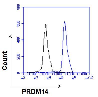
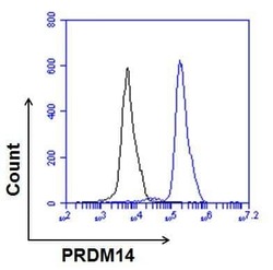
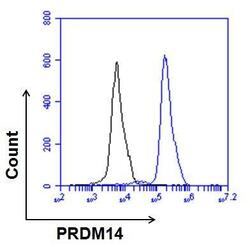
- Flow cytometry analysis of PRDM14 on human Gibco® Episomal iPSC line (Product # A18945) grown on vitronectin in Essential 8 medium (Product # A1517001). Cells were fixed, permeabilized, and then stained with 5% FBS/PBS (black histogram) or 5 µg/mL of PRDM14 rabbit polyclonal antibody (Product # PA1-114, blue histogram) in 5% FBS/PBS. After incubation of the primary antibody for 1 hour on ice, the cells were stained with a DyLight 488-conjugated goat anti-rabbit (Product # 35553) at a dilution of 1:500 for 1 hour on ice. A representative 10,000 cells were acquired for each sample.
- Submitted by
- Invitrogen Antibodies (provider)
- Main image
- Experimental details
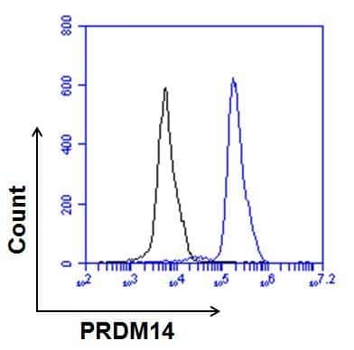
- Flow cytometry analysis of PRDM14 on human Gibco® Episomal iPSC line (Product # A18945) grown on vitronectin in Essential 8 medium (Product # A1517001). Cells were fixed, permeabilized, and then stained with 5% FBS/PBS (black histogram) or 5 µg/mL of PRDM14 rabbit polyclonal antibody (Product # PA1-114, blue histogram) in 5% FBS/PBS. After incubation of the primary antibody for 1 hour on ice, the cells were stained with a DyLight 488-conjugated goat anti-rabbit (Product # 35553) at a dilution of 1:500 for 1 hour on ice. A representative 10,000 cells were acquired for each sample.