 Explore
 Validate
 Learn
Learn Immunoprecipitation
 Flow cytometry
Antibody data
- Antibody Data
- Antigen structure
- References [5]
- Comments [0]
- Validations
- Flow cytometry [1]
- Other assay [1]
Submit
Validation data
Reference
Comment
Report error
- Product number
- 14-0496-82 - Provider product page

- Provider
- Invitrogen Antibodies
- Product name
- CD49e (Integrin alpha 5) Monoclonal Antibody (eBioSAM-1 (SAM-1, SAM1)), eBioscience™
- Antibody type
- Monoclonal
- Antigen
- Other
- Description
- Description: The eBioSAM-1 monoclonal antibody reacts with human integrin alpha 5, also known as fibronectin receptor alpha chain, very late activation antigen 5 alpha, and CD49e. Integrins are composed of an alpha chain and a beta chain, which non-covalently associate to form the functional integrin. Integrin heterodimers participate in cell surface-mediated signaling and adhesion functions. Integrin alpha 5 undergoes post-translational cleavage in its extracellular domain to yield disulfide linked light and heavy chains that join with Integrin beta 1 (CD29) to form the fibronectin receptor, also known as the very late activation antigen-5 (VLA-5) complex. Integrin alpha 5 is expressed on thymocytes, T cells, monocytes, platelets, early B cells, and activated B cells. Applications Reported: This eBioSAM-1 (SAM-1, SAM1) antibody has been reported for use in flow cytometric analysis, immunoprecipitation, and immunohistochemical staining. Applications Tested: This eBioSAM-1 (SAM-1, SAM1) antibody has been tested by flow cytometric analysis of normal human peripheral blood cells. This can be used at less than or equal to 0.25 µg per test. A test is defined as the amount (µg) of antibody that will stain a cell sample in a final volume of 100 µL. Cell number should be determined empirically but can range from 10^5 to 10^8 cells/test. It is recommended that the antibody be carefully titrated for optimal performance in the assay of interest. Purity: Greater than 90%, as determined by SDS-PAGE. Aggregation: Less than 10%, as determined by HPLC. Filtration: 0.2 µm post-manufacturing filtered.
- Reactivity
- Human
- Host
- Mouse
- Isotype
- IgG
- Antibody clone number
- eBioSAM-1 (SAM-1, SAM1)
- Vial size
- 100 µg
- Concentration
- 0.5 mg/mL
- Storage
- 4° C
Submitted references Human SND2 mediates ER targeting of GPI-anchored proteins with low hydrophobic GPI attachment signals.
Porcine mononuclear cells adhere to human fibronectin independently of very late antigen-5: implications for donor-specific tolerance induction in xenotransplantation.
Monocyte-derived dendritic cells have a phenotype comparable to that of dermal dendritic cells and display ultrastructural granules distinct from Birbeck granules.
Signal transduction through the beta1 integrin family surface adhesion molecules VLA-4 and VLA-5 of human B-cell precursors activates CD19 receptor-associated protein-tyrosine kinases.
An alpha 5 beta 1-like integrin receptor mediates the binding of less pathogenic Candida species to fibronectin.
Yang J, Hirata T, Liu YS, Guo XY, Gao XD, Kinoshita T, Fujita M
FEBS letters 2021 Jun;595(11):1542-1558
FEBS letters 2021 Jun;595(11):1542-1558
Porcine mononuclear cells adhere to human fibronectin independently of very late antigen-5: implications for donor-specific tolerance induction in xenotransplantation.
Theodore PR, Simon AR, Warrens AN, Sackstein R, Sykes M
Xenotransplantation 2002 Jul;9(4):277-89
Xenotransplantation 2002 Jul;9(4):277-89
Monocyte-derived dendritic cells have a phenotype comparable to that of dermal dendritic cells and display ultrastructural granules distinct from Birbeck granules.
Grassi F, Dezutter-Dambuyant C, McIlroy D, Jacquet C, Yoneda K, Imamura S, Boumsell L, Schmitt D, Autran B, Debré P, Hosmalin A
Journal of leukocyte biology 1998 Oct;64(4):484-93
Journal of leukocyte biology 1998 Oct;64(4):484-93
Signal transduction through the beta1 integrin family surface adhesion molecules VLA-4 and VLA-5 of human B-cell precursors activates CD19 receptor-associated protein-tyrosine kinases.
Xiao J, Messinger Y, Jin J, Myers DE, Bolen JB, Uckun FM
The Journal of biological chemistry 1996 Mar 29;271(13):7659-64
The Journal of biological chemistry 1996 Mar 29;271(13):7659-64
An alpha 5 beta 1-like integrin receptor mediates the binding of less pathogenic Candida species to fibronectin.
Santoni G, Birarelli P, Hong LJ, Gamero A, Djeu JY, Piccoli M
Journal of medical microbiology 1995 Nov;43(5):360-7
Journal of medical microbiology 1995 Nov;43(5):360-7
No comments: Submit comment
Supportive validation
- Submitted by
- Invitrogen Antibodies (provider)
- Main image
- Experimental details
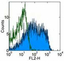
- Staining of normal human peripheral blood cells with 0.25 µg of Mouse IgG1 kappa Isotype Control Purified (Product # 14-4714-82) (open histogram) or 0.25 µg of Anti-Human CD49e (Integrin alpha 5) Purified (filled histogram) followed by Anti-Mouse IgG Biotin (Product # 13-4013-85) and Streptavidin PE (Product # 12-4317-87). Cells in the lymphocyte gate were used for analysis.

Supportive validation
- Submitted by
- Invitrogen Antibodies (provider)
- Main image

- Experimental details
- 1 Fig. Knockout of hSND2 causes a reduction in GPI-AP expression. (A) Schematic representation of the hSND2 protein. The regions corresponding to the gene deletion using CRISPR/Cas9 systems are shown as black arrows. Two sgRNA sequences were used for knocking out hSND2. TM, transmembrane domain. (B) Confirmation of hSND2 gene KO. Genomic DNA was extracted from wild-type and hSND2-KO No. 5 clonal cell lines, and a DNA fragment was amplified around the KO region. A decreased DNA band was detected in the hSND2-KO cells compared with the wild-type cells. The expected size of the DNA fragment for hSND2 is 206 bp in wild-type cells and 135 bp in KO cells. The 71-bp sequence was deleted in the hSND2-KO No. 5 cell line. Capital bold letters, the target sequences; underlined letters, the PAM sequence; dotted lines, the deletion sequence. WT, wild-type cells; KO, knockout cells. (C) Flow cytometric analysis of the CD59, DAF, CD109, BSG, and CD49e proteins in HEK293 and hSND2-KO cells. The cells were stained with appropriate antibodies. Black solid lines, each protein's staining in HEK293 cells; gray shaded areas, each protein's staining in hSND2-KO cells; dashed black lines, HEK293 cells only stained with second antibody (background). (D) Analysis and graphical representation of C. Quantitative data of the mean fluorescence intensity of three independent experiments are shown as the means +- SD. The value in HEK293 cells was set as 1, and the relative values in hSND2-KO cells are shown
