Explore
 Validate
 Learn
Learn Western blot
Antibody data
- Antibody Data
- Antigen structure
- References [1]
- Comments [0]
- Validations
- Western blot [1]
- Flow cytometry [1]
- Other assay [2]
Submit
Validation data
Reference
Comment
Report error
- Product number
- PA5-25433 - Provider product page

- Provider
- Invitrogen Antibodies
- Product name
- ITGA5 Polyclonal Antibody
- Antibody type
- Polyclonal
- Antigen
- Synthetic peptide
- Reactivity
- Human, Mouse
- Host
- Rabbit
- Isotype
- IgG
- Vial size
- 400 µL
- Concentration
- 0.5 mg/mL
- Storage
- -20° C, Avoid Freeze/Thaw Cycles
Submitted references MicroRNA-26a inhibits wound healing through decreased keratinocytes migration by regulating ITGA5 through PI3K/AKT signaling pathway.
Jiang Z, Wei J, Yang W, Li W, Liu F, Yan X, Yan X, Hu N, Li J
Bioscience reports 2020 Sep 30;40(9)
Bioscience reports 2020 Sep 30;40(9)
No comments: Submit comment
Supportive validation
- Submitted by
- Invitrogen Antibodies (provider)
- Main image

- Experimental details
- Western blot analysis in mouse bladder tissue lysates (15 µg per lane) using an ITGA5 polyclonal antibody (Product # PA5-25433).

Supportive validation
- Submitted by
- Invitrogen Antibodies (provider)
- Main image

- Experimental details
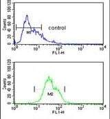
- Flow cytometry analysis of CEM cells using an ITGA5 polyclonal antibody (Product # PA5-25433) (bottom) compared to a negative control cell (top) at a dilution of 1:10-50, followed by a FITC-conjugated goat anti-rabbit antibody
Supportive validation
- Submitted by
- Invitrogen Antibodies (provider)
- Main image
- Experimental details
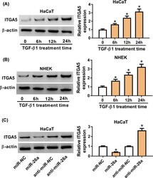
- Figure 4 ITGA5 was up-regulated by TGF-beta1 and modulated by miR-26a ( A ) HaCaT cells were treated with 2 ng/mL TGF-beta1 for 0, 6, 12 and 24 h. The ITGA5 expression in the treated HaCaT cells was detected by Western blot assay. ( B ) ITGA5 protein expression of NHEK after 2 ng/mL TGF-beta1 treatment for 0, 6, 12 and 24 h was detected by Western blot assay. ( C ) ITGA5 protein expression in HaCaT cells after transfection of miR-26a inhibitor or miR-26a was measured by Western blot assay. beta-actin was used as reference for ITGA5. * means p -value less than 0.05.

- Submitted by
- Invitrogen Antibodies (provider)
- Main image

- Experimental details
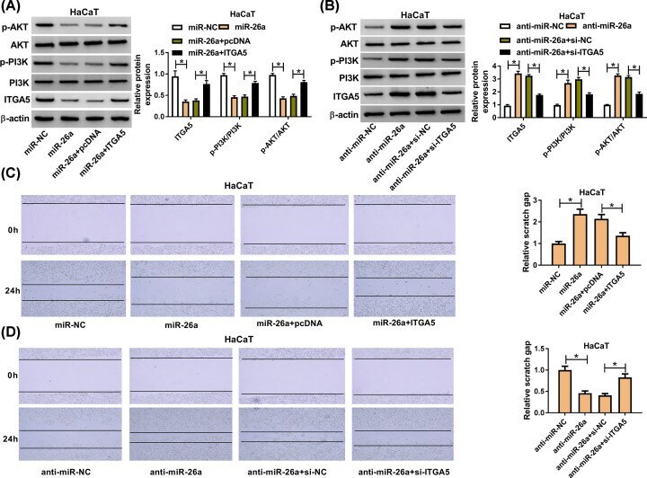
- Figure 5 ITGA5 reversed the effect of miR-26a on cell migration via PI3K/AKT signaling pathway ( A ) The expression level of ITGA5 was detected by Western blot assay after transfection with miR-26a or miR-26a+ITGA5 in HaCaT cells. ( B ) The expression level of ITGA5, PI3K and AKT was measured after transfection with anti-miR-26a or anti-miR-26a+si-ITGA5. ( C,D ) The migration ability of HaCaT cell was analyzed after overexpressing both miR-26a and ITGA5 in HaCaT cells after TGF-beta1 treatment for 24 h. (B) The migration cell number of HaCaT after transfection of miR-26a inhibitor and si-ITGA5 after TGF-beta1 treatment for 24 h. * means P -value less than 0.05.