Explore
 Validate
 Learn
Learn Western blot
Antibody data
- Antibody Data
- Antigen structure
- References [0]
- Comments [0]
- Validations
- Western blot [4]
- Immunocytochemistry [3]
- Immunohistochemistry [2]
Submit
Validation data
Reference
Comment
Report error
- Product number
- MA5-17279 - Provider product page

- Provider
- Invitrogen Antibodies
- Product name
- COX4 Monoclonal Antibody (GT6310)
- Antibody type
- Monoclonal
- Antigen
- Recombinant protein fragment
- Description
- Recommended positive controls: 293T, A431, HeLa, HepG2, A375, rat muscle.
- Antibody clone number
- GT6310
- Concentration
- 1 mg/mL
No comments: Submit comment
Supportive validation
- Submitted by
- Invitrogen Antibodies (provider)
- Main image
- Experimental details
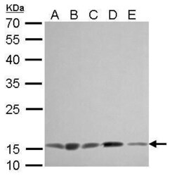
- COX4 Monoclonal Antibody (GT6310) detects COX4I1 protein by Western blot analysis. A. 30 µg 293T whole cell lysate/extract. B. 30 µg A431 whole cell lysate/extract. C. 30 µg HeLa whole cell lysate/extract. D. 30 µg HepG2 whole cell lysate/extract. E. 30 µg A375 whole cell lysate/extract.12 % SDS-PAGE. COX4 Monoclonal Antibody (GT6310) (Product # MA5-17279) dilution: 1:1,000.

- Submitted by
- Invitrogen Antibodies (provider)
- Main image

- Experimental details
- COX4 Monoclonal Antibody (GT6310) detects COX4I1 protein by Western blot analysis. A. 50 µg rat muscle lysate/extract.12 % SDS-PAGE. COX4 Monoclonal Antibody (GT6310) (Product # MA5-17279) dilution: 1:1,000.

- Submitted by
- Invitrogen Antibodies (provider)
- Main image

- Experimental details
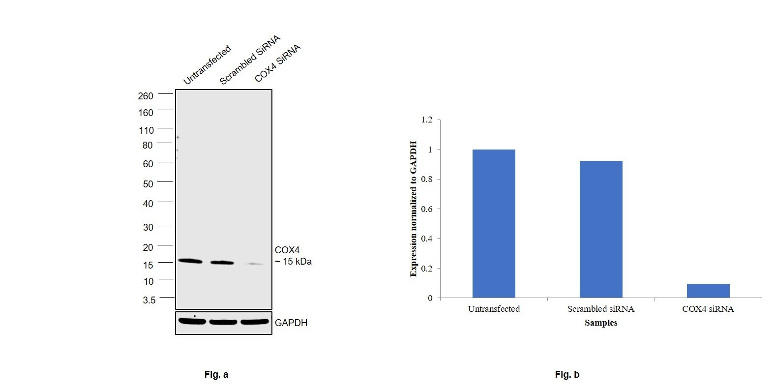
- Knockdown of COX4 was achieved by transfecting Hep G2 with COX4 specific siRNAs (Silencer® select Product # S3386, S3387). Western blot analysis (Fig. a) was performed using Whole cell extracts from the COX4 knockdown cells (lane 3), non-targeting scrambled siRNA transfected cells (lane 2) and untransfected cells (lane 1). The blot was probed with COX4 Monoclonal Antibody (GT6310) (Product # MA5-17279, 1:1000 dilution) and Goat anti-Mouse IgG (H+L) Superclonal™ Recombinant Secondary Antibody, HRP (Product # A28177, 1:4000 dilution). Densitometric analysis of this western blot is shown in histogram (Fig. b). Decrease in signal upon siRNA mediated knock down confirms that antibody is specific to COX4.
- Submitted by
- Invitrogen Antibodies (provider)
- Main image
- Experimental details
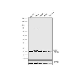
- Western blot was performed using Anti-COX4 Monoclonal Antibody (GT6310)(Product # MA5-17279) and a 15kDa band corresponding to COX4 was observed across the cell lines and tissues tested. Whole cell extracts (30 µg lysate) of Hep G2 (Lane 1), HeLa (Lane 2), LNCaP (Lane 3), A-431 (Lane 4) and Rat Heart (Lane 5) were electrophoresed using NuPAGE™ 4-12% Bis-Tris Protein Gel (Product # NP0321BOX). Resolved proteins were then transferred onto a Nitrocellulose membrane (Product # LC2001) by iBlot® 2 Dry Blotting System (Product # IB21001). The blot was probed with the primary antibody (1:1000 dilution) and detected by chemiluminescence with Goat anti-Mouse IgG (H+L) Superclonal™ Recombinant Secondary Antibody, HRP (Product # A28177, 1:4000 dilution) using the iBright FL 1000 (Product # A32752). Chemiluminescent detection was performed using Novex® ECL Chemiluminescent Substrate Reagent Kit (Product # WP20005).

Supportive validation
- Submitted by
- Invitrogen Antibodies (provider)
- Main image

- Experimental details
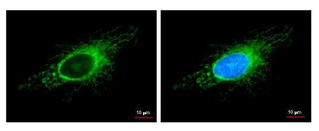
- COX4I1 antibody detects COX4I1 protein at mitochondria by immunofluorescent analysis. Sample: HeLa cells were fixed in -20ºC 100% methanol for 5 min. Green: COX4I1 protein stained by COX4I1 antibody (Product # MA5-17279) diluted at 1:500. Blue: Hoechst 33342 staining.

- Submitted by
- Invitrogen Antibodies (provider)
- Main image

- Experimental details
- COX4I1 antibody detects COX4I1 protein at mitochondria by immunofluorescent analysis. Sample: HeLa cells were fixed in -20ºC 100% methanol for 5 min. Green: COX4I1 protein stained by COX4I1 antibody (Product # MA5-17279) diluted at 1:500. Blue: Hoechst 33342 staining.
- Submitted by
- Invitrogen Antibodies (provider)
- Main image

- Experimental details
- Immunofluorescence analysis of COX4 was performed using 70% confluent log phase Hep G2 cells. The cells were fixed with 4% paraformaldehyde for 10 minutes, permeabilized with 0.1% Triton™ X-100 for 10 minutes, and blocked with 2% BSA for 45 minutes at room temperature. The cells were labeled with COX4 Monoclonal Antibody (GT6310) (Product # MA5-17279) at 1:100 dilution in 0.1% BSA, incubated at 4 degree celsius overnight and then labeled with Donkey anti-Mouse IgG (H+L) Highly Cross-Adsorbed Secondary Antibody, Alexa Fluor Plus 647 (Product # A32787), (1:2000 dilution), for 45 minutes at room temperature (Panel a: Green). Nuclei (Panel b:Blue) were stained with SlowFade® Gold Antifade Mountant with DAPI (Product # S36938). F-actin (Panel c: Red) was stained with Rhodamine Phalloidin (Product # R415, 1:300 dilution). Panel d represents the merged image showing Mitochondrial localization. Panel e represents control cells with no primary antibody to assess background. The images were captured at 60X magnification.

Supportive validation
- Submitted by
- Invitrogen Antibodies (provider)
- Main image

- Experimental details
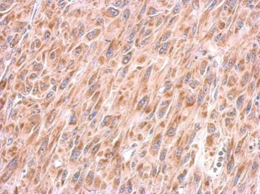
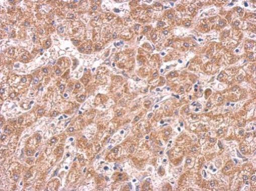
- COX4 Monoclonal Antibody (GT6310) detects COX4I1 protein at cytosol on human hepatoma by immunohistochemical analysis. Sample: Paraffin-embedded hepatoma. COX4 Monoclonal Antibody (GT6310) (Product # MA5-17279) dilution: 1:200. Antigen Retrieval: EDTA based buffer, pH 8.0, 15 min.
- Submitted by
- Invitrogen Antibodies (provider)
- Main image

- Experimental details
- COX4 Monoclonal Antibody (GT6310) detects COX4I1 protein at cytosol on U87 xenograft by immunohistochemical analysis. Sample: Paraffin-embedded U87 xenograft. COX4 Monoclonal Antibody (GT6310) (Product # MA5-17279) dilution: 1:200. Antigen Retrieval: EDTA based buffer, pH 8.0, 15 min.