 Explore
 Validate
 Learn
LearnSTJ99044
antibody from St John's Laboratory
Targeting: COX4I1
COX4, COX4-1
 Western blot Immunocytochemistry
 Immunoprecipitation Immunohistochemistry Flow cytometry Chromatin Immunoprecipitation Other assay
Antibody data
- Antibody Data
- Antigen structure
- References [0]
- Comments [0]
- Validations
- Western blot [2]
- Immunocytochemistry [1]
- Immunohistochemistry [1]
- Chromatin Immunoprecipitation [1]
- Other assay [1]
Submit
Validation data
Reference
Comment
Report error
- Product number
- STJ99044 - Provider product page

- Provider
- St John's Laboratory
- Product name
- Anti-COX4I1 antibody [4D11-B3-E8] (STJ99044)
- Antibody type
- Monoclonal
- Description
- Mouse monoclonal antibody anti-Cytochrome C Oxidase Subunit 4 Isoform 1-Mitochondrial is suitable for use in Western Blot, Flow Cytometry, Immunocytochemistry, Immunoprecipitation and Immunohistochemistry research applications.
- Reactivity
- Human, Mouse, Rat, Goat, Hamster, Simian
- Host
- Mouse
- Conjugate
- Unconjugated
- Antigen sequence
NA- Epitope
- NA
- Isotype
- IgG
- Antibody clone number
- NA
- Vial size
- NA
- Concentration
- NA
- Storage
- Store at-20°C for up to 1 year from the date of receipt, and avoid repeat freeze-thaw cycles.
- Handling
- NA
No comments: Submit comment
Supportive validation
Supportive validation
- Submitted by
- St John's Laboratory (provider)
- Main image

- Experimental details
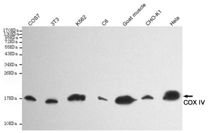
- Western blot detection of COX IV in Goat muscle, CHO-k1, COS7, 3T3, Hela, C6 and K562 cell lysates using COX IV mouse mAb (1:5000 diluted).Predicted band size:17KDa.Observed band size:17KDa.
- Sample type
- NA
- Validation comment
- NA
- Primary Ab dilution
- NA
- Other comments
- NA
- Secondary Ab
- NA
- Secondary Ab dilution
- NA
- Protocol
- NA
Supportive validation
- Submitted by
- St John's Laboratory (provider)
- Main image
- Experimental details
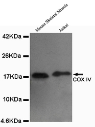
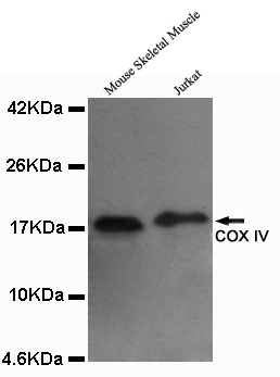
- Western blot detection of COX IV in Mouse skeletal muscel and Jurkat lysates using COX IV mouse mAb (1:1000 diluted). Predicted band size: 17KDa.Observed band size: 17KDa.
- Sample type
- NA
- Validation comment
- NA
- Primary Ab dilution
- NA
- Other comments
- NA
- Secondary Ab
- NA
- Secondary Ab dilution
- NA
- Protocol
- NA
Supportive validation
- Submitted by
- St John's Laboratory (provider)
- Main image

- Experimental details
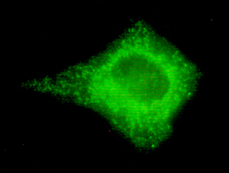
- Immunocytochemistry of HeLa cells using anti-COX IV mouse mAb diluted 1:150.
- Sample type
- NA
- Validation comment
- NA
- Primary Ab dilution
- NA
- Other comments
- NA
- Secondary Ab
- NA
- Secondary Ab dilution
- NA
- Protocol
- NA
Supportive validation
- Submitted by
- St John's Laboratory (provider)
- Main image

- Experimental details
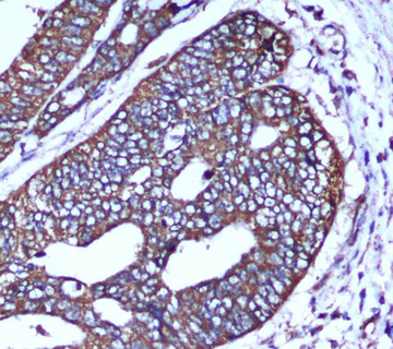
- Immunohistochemical analysis of paraffin-embedded human colorectal carcinoma with COX IV Mouse mAb (4D11-B3-E8, 1:50 diluted) , showing cytoplasm localization.A high pressure mediated antigen retrieval step was performed in citrate buffer (pH6.0).
- Sample type
- NA
- Validation comment
- NA
- Primary Ab dilution
- NA
- Other comments
- NA
- Secondary Ab
- NA
- Secondary Ab dilution
- NA
- Protocol
- NA
Supportive validation
- Submitted by
- St John's Laboratory (provider)
- Main image

- Experimental details
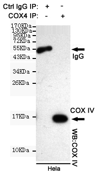
- Immunoprecipitation analysis of Hela cell lysates using COX IV mouse mAb.
- Sample type
- NA
- Validation comment
- NA
- Primary Ab dilution
- NA
- Other comments
- NA
- Secondary Ab
- NA
- Secondary Ab dilution
- NA
- Protocol
- NA
Supportive validation
- Submitted by
- St John's Laboratory (provider)
- Main image
- Experimental details
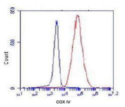
- Flow Cytometry analysis of K562 cells stained with COX4 (red, 1/100 dilution) , followed by FITC-conjugated goat anti-mouse IgG. Blue line histogram represents the isotype control, normal mouse IgG.
- Sample type
- NA
- Validation comment
- NA
- Primary Ab dilution
- NA
- Other comments
- NA
- Secondary Ab
- NA
- Secondary Ab dilution
- NA
- Protocol
- NA
