Explore
 Validate
 Learn
LearnMA5-15078
antibody from Invitrogen Antibodies
Targeting: COX4I1
COX4, COX4-1
 Western blot Immunocytochemistry
 Immunoprecipitation Immunohistochemistry Flow cytometry Other assay
Antibody data
- Antibody Data
- Antigen structure
- References [7]
- Comments [0]
- Validations
- Western blot [3]
- Immunocytochemistry [2]
- Immunohistochemistry [1]
- Other assay [6]
Submit
Validation data
Reference
Comment
Report error
- Product number
- MA5-15078 - Provider product page

- Provider
- Invitrogen Antibodies
- Product name
- COX4 Monoclonal Antibody (K.473.4)
- Antibody type
- Monoclonal
- Antigen
- Synthetic peptide
- Description
- It is not recommended to aliquot this antibody.
- Reactivity
- Human, Rat, Bovine, Porcine, Zebrafish
- Host
- Rabbit
- Isotype
- IgG
- Antibody clone number
- K.473.4
- Vial size
- 100 μL
- Concentration
- 331 μg/mL
- Storage
- -20°C
Submitted references Cardiac disruption of SDHAF4-mediated mitochondrial complex II assembly promotes dilated cardiomyopathy.
Interspecies Differences in Proteome Turnover Kinetics Are Correlated With Life Spans and Energetic Demands.
Abnormal expression of lncRNA UCA1 disturbed cell apoptosis through mediating mitochondrial dynamics in PDAC.
On the potential role of globins in brown adipose tissue: a novel conceptual model and studies in myoglobin knockout mice.
PARK14 PLA2G6 mutants are defective in preventing rotenone-induced mitochondrial dysfunction, ROS generation and activation of mitochondrial apoptotic pathway.
3,5-Diiodo-L-thyronine activates brown adipose tissue thermogenesis in hypothyroid rats.
A nuclear role for the respiratory enzyme CLK-1 in regulating mitochondrial stress responses and longevity.
Wang X, Zhang X, Cao K, Zeng M, Fu X, Zheng A, Zhang F, Gao F, Zou X, Li H, Li M, Lv W, Xu J, Long J, Zang W, Chen J, Gao F, Ding J, Liu J, Feng Z
Nature communications 2022 Jul 8;13(1):3947
Nature communications 2022 Jul 8;13(1):3947
Interspecies Differences in Proteome Turnover Kinetics Are Correlated With Life Spans and Energetic Demands.
Swovick K, Firsanov D, Welle KA, Hryhorenko JR, Wise JP Sr, George C, Sformo TL, Seluanov A, Gorbunova V, Ghaemmaghami S
Molecular & cellular proteomics : MCP 2021;20:100041
Molecular & cellular proteomics : MCP 2021;20:100041
Abnormal expression of lncRNA UCA1 disturbed cell apoptosis through mediating mitochondrial dynamics in PDAC.
Teng B, Feng T, Li W, Wang Z
Neoplasma 2021 Mar;68(2):334-341
Neoplasma 2021 Mar;68(2):334-341
On the potential role of globins in brown adipose tissue: a novel conceptual model and studies in myoglobin knockout mice.
Blackburn ML, Wankhade UD, Ono-Moore KD, Chintapalli SV, Fox R, Rutkowsky JM, Willis BJ, Tolentino T, Lloyd KCK, Adams SH
American journal of physiology. Endocrinology and metabolism 2021 Jul 1;321(1):E47-E62
American journal of physiology. Endocrinology and metabolism 2021 Jul 1;321(1):E47-E62
PARK14 PLA2G6 mutants are defective in preventing rotenone-induced mitochondrial dysfunction, ROS generation and activation of mitochondrial apoptotic pathway.
Chiu CC, Yeh TH, Lu CS, Huang YC, Cheng YC, Huang YZ, Weng YH, Liu YC, Lai SC, Chen YL, Chen YJ, Chen CL, Chen HY, Lin YW, Wang HL
Oncotarget 2017 Oct 3;8(45):79046-79060
Oncotarget 2017 Oct 3;8(45):79046-79060
3,5-Diiodo-L-thyronine activates brown adipose tissue thermogenesis in hypothyroid rats.
Lombardi A, Senese R, De Matteis R, Busiello RA, Cioffi F, Goglia F, Lanni A
PloS one 2015;10(2):e0116498
PloS one 2015;10(2):e0116498
A nuclear role for the respiratory enzyme CLK-1 in regulating mitochondrial stress responses and longevity.
Monaghan RM, Barnes RG, Fisher K, Andreou T, Rooney N, Poulin GB, Whitmarsh AJ
Nature cell biology 2015 Jun;17(6):782-92
Nature cell biology 2015 Jun;17(6):782-92
No comments: Submit comment
Supportive validation
- Submitted by
- Invitrogen Antibodies (provider)
- Main image

- Experimental details
- Western blot analysis of COX IV in extracts from HeLa and Jurkat cell lines using COX IV monoclonal antibody (Product # MA5-15078).

- Submitted by
- Invitrogen Antibodies (provider)
- Main image
- Experimental details
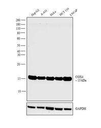
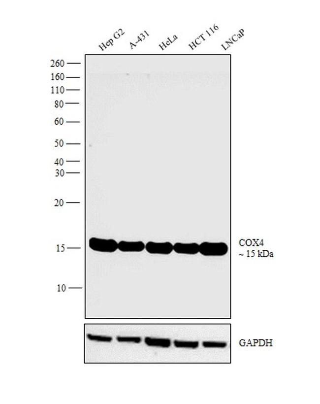
- Western blot analysis was performed on whole cell extracts (30 µg lysate) of Hep G2 (Lane 1), A-431 (Lane 2), HeLa (Lane 3), HCT 116 (Lane 4) and LNCaP (Lane 5). The blot was probed with COX4 Monoclonal Antibody (Product # MA5-15078, 1:1,000 dilution) and detected by chemiluminescence using Goat anti-Rabbit IgG (Heavy Chain) Superclonal™ Secondary Antibody, HRP conjugate (Product # A27036, 0.25 µg/mL, 1:4,000 dilution). A band at 15 kDa corresponding to COX4 was observed across the cell lines tested.
- Submitted by
- Invitrogen Antibodies (provider)
- Main image
- Experimental details
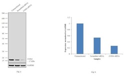
- Knockdown of COX4 was achieved by transfecting Hep G2 cells with COX4 specific siRNAs (Silencer® select Product # s3388). Western blot analysis (Fig. a) was performed using whole cell extracts from the COX4 knockdown cells (lane 3), non-specific scrambled siRNA transfected cells (lane 2) and untransfected cells (lane 1). The blots were probed with COX4 Monoclonal Antibody (Product # MA5-15078, 1:1,000 dilution) and Goat anti-Rabbit IgG (Heavy Chain) Superclonal™ Secondary Antibody, HRP conjugate (Product # A27036, 0.25 µg/mL, 1:4,000 dilution). Densitometric analysis of this western blot is shown in histogram (Fig. b). Decrease in signal upon siRNA mediated knock down confirms that antibody is specific to COX4.

Supportive validation
- Submitted by
- Invitrogen Antibodies (provider)
- Main image

- Experimental details
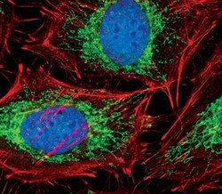
- Immunofluorescent analysis of COX IV in HeLa cells using a COX IV monoclonal antibody (Product # MA5-15078) (green). Actin filaments are labeled with a fluorescent red phalloidin. DNA is labeled using a fluorescent blue dye.

- Submitted by
- Invitrogen Antibodies (provider)
- Main image
- Experimental details
- Immunofluorescent analysis of COX IV in HeLa cells using a COX IV monoclonal antibody (Product # MA5-15078) (green). Actin filaments are labeled with a fluorescent red phalloidin. DNA is labeled using a fluorescent blue dye.

Supportive validation
- Submitted by
- Invitrogen Antibodies (provider)
- Main image
- Experimental details
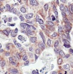
- Immunohistochemical analysis of COX IV in paraffin-embedded human colon carcinoma using a COX IV monoclonal antibody (Product # MA5-15078) showing staining of the mitochondria.

Supportive validation
- Submitted by
- Invitrogen Antibodies (provider)
- Main image
- Experimental details
- NULL
- Submitted by
- Invitrogen Antibodies (provider)
- Main image
- Experimental details
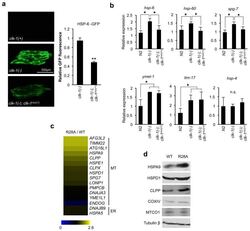
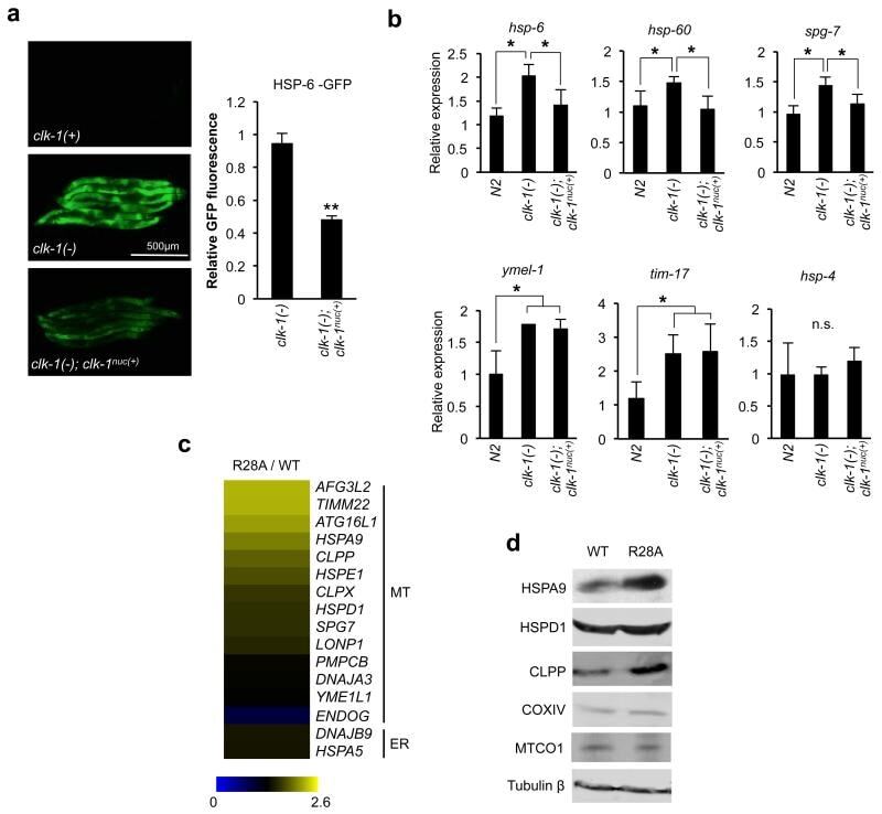
- Figure 6 Nuclear CLK-1/COQ7 suppresses the expression of a subset of UPR mt genes (a) CLK-1 nuc(+) expressing worms display decreased hsp-6::gfp reporter activity compared to clk-1(-) worms. The unmodified hsp-6::gfp reporter strain is designated clk-1(+) . Quantification of reporter fluorescence in CLK-1 nuc(+) expressing worms ( clk-1(-); clk-1 nuc(+) ) relative to clk-1(-) worms (mean fluorescence of 50 worms per genotype pooled from n=3 independent experiments; error bars, s.e.m. ** P < 0.005). (b) qPCR measuring mRNA transcripts of UPR mt genes in clk-1(-) or clk-1(-); clk-1 nuc(+) worms relative to wild type strain ( N2 ) (mean values from 3 reactions per condition in n=3 independent experiments; error bars, s.e.m., n.s., no significant difference, * P < 0.05). (c) Heat map depicting change in expression of UPR mt genes (MT) and UPR ER (ER) genes in R28A cells compared to WT COQ7 cells. Map generated from the qPCR data presented in Supplementary Figure 4b and is representative of n=3 independent experiments. Scale represents mean fold change in expression. (d) Immunoblots of levels of UPR mt proteins including the mitochondrial controls COXIV (nuclear-encoded) and MTCO1 (mitochondrial-encoded). Uncropped images of blots are shown in Supplementary Fig. 5 .
- Submitted by
- Invitrogen Antibodies (provider)
- Main image
- Experimental details
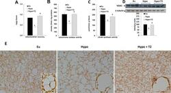
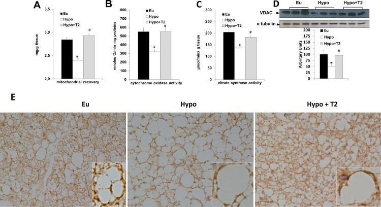
- Figure 5 Effects of hypothyroidism and T2 administration to Hypo rats on tissue mitochondrial content and oxidative capacity. (A) Mitochondrial protein/100 mg tissue, (B) cytochrome oxidase (COX) activity, (C) citrate synthase activity, and (D) VDAC1 content detected in whole BAT lysates. Data in the bar chart are represented as means +- standard error of the mean from five independent experiments, each performed in duplicate. *P
- Submitted by
- Invitrogen Antibodies (provider)
- Main image
- Experimental details
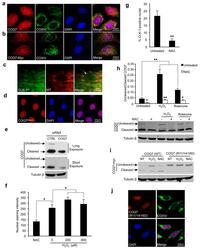
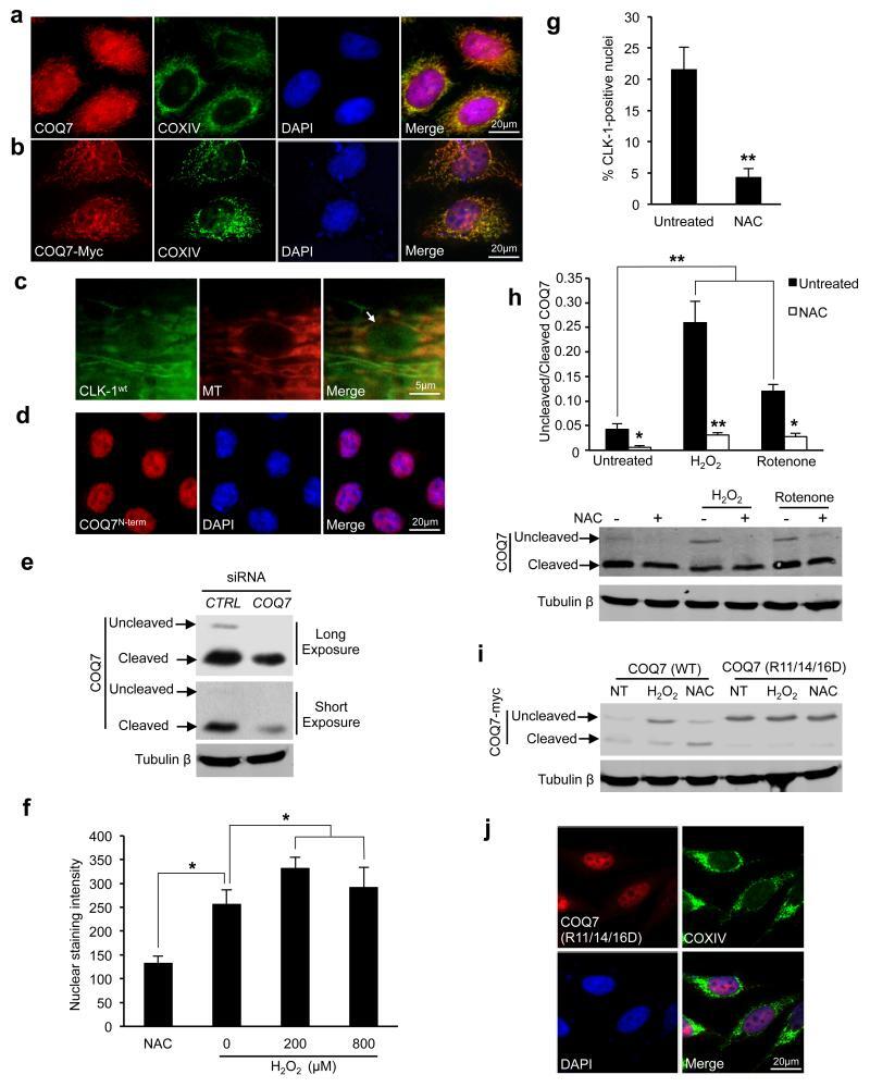
- Figure 1 CLK-1 and its human homologue COQ7 localise to mitochondria and nuclei ( a ) Endogenous COQ7 and COXIV (mitochondrial marker) immunostaining in HeLa cells. Nuclei stained with DAPI. ( b ) HeLa cells expressing COQ7 tagged at the C-terminus with Myc epitope (COQ7-Myc) were stained with anti-Myc and anti-COXIV antibodies. Quantification of nuclear COQ7-Myc staining is in Supplementary Fig. 1a . ( c ) Wild type CLK-1 (CLK-1 wt ) fused at the C-terminus to GFP localises to both mitochondria and nuclei in adult C. elegans . Arrow marks nucleus. MT (MitoTracker, mitochondrial marker). ( d ) HeLa cells immunostained with an antibody specific to the N-terminus (amino acids 1-37) of COQ7 (COQ7 N-term ). (e) siRNA targeting COQ7 transcripts decrease levels of both cleaved and uncleaved COQ7 protein. Immunoblots of lysates from HEK293 cells transfected with non-targeting ( CTRL ) or COQ7 siRNA. Short and long exposures are shown. ( f ) Quantification of nuclear staining intensity of cells immunostained with anti-COQ7(1-37) (COQ7 N-term ) following treatment with antioxidant (N-acetyl cysteine, NAC, 10 mM, 24 h) or exogenous ROS (hydrogen peroxide, 200 muM or 800 muM, respectively, 3 h) compared to untreated (0) control. 50 cells assessed per experiment in n=3 independent experiments (error bars, s.e.m. * P < 0.05). (g) Percent of mCherry-positive nuclei that are also GFP-positive in C. elegans expressing CLK-1-GFP and the nuclear marker HIS-24-mCherry. 25 worms were assessed pe
- Submitted by
- Invitrogen Antibodies (provider)
- Main image

- Experimental details
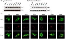
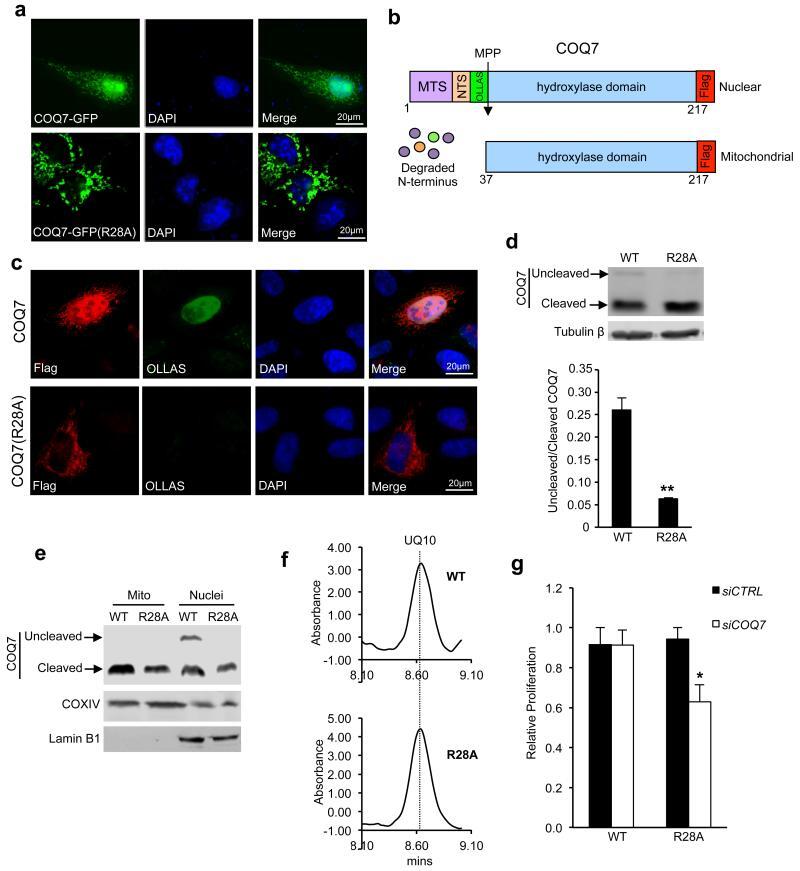
- Figure 2 Nuclear COQ7 functions independently of the mitochondrial form (a) Reduced nuclear localisation of COQ7 (R28A) mutant. Fluorescence in COS7 cells expressing COQ7 or COQ7 (R28A) fused at the C-terminus to GFP. Nuclei stained with DAPI. (b) Schematic depicting location of the mitochondrial targeting sequence (MTS), the nuclear targeting sequence (NTS) and the mitochondrial processing peptidase cleavage site (MPP) on COQ7. The N-terminal region of COQ7 is degraded following cleavage by MPP in mitochondria. Also shown are the positions of the OLLAS and FLAG epitope-tags. Numbers refer to amino acid positions of COQ7. ( c ) HeLa cells expressing dual OLLAS and FLAG tagged COQ7 or COQ7 (R28A) immunostained with anti-FLAG and anti-OLLAS antibodies. The anti-FLAG antibodies recognise total COQ7 (uncleaved and cleaved) and the anti-OLLAS antibody specifically recognises uncleaved nuclear COQ7. (d) Immunoblot of cell lysates from HEK293 cells stably expressing either untagged wild type (WT) COQ7 or the R28A mutant in the presence of siRNA against untranslated regions of COQ7 . Quantification of the ratio of uncleaved (nuclear) to cleaved (mitochondrial) COQ7 from n=3 independent experiments is presented (error bars, s.e.m. ** P < 0.005). (e) Immunoblot of cell lysates from the stable HEK293 cells expressing either wild type (WT) COQ7 or the R28A mutant separated into mitochondrial (Mito) and nuclear (Nuclei) pellet fractions (COXIV, mitochondrial marker; Lamin B1, nuclear matr
- Submitted by
- Invitrogen Antibodies (provider)
- Main image
- Experimental details
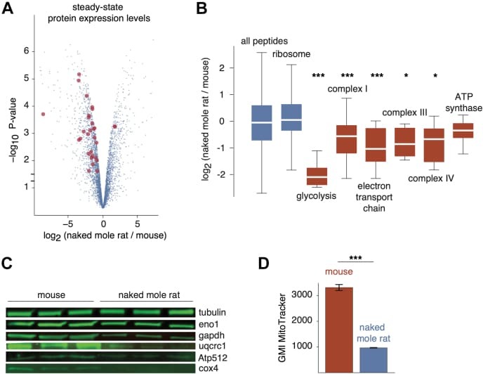
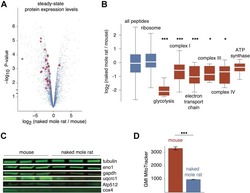
- Fig. 5 Proteome-wide differences in steady-state protein levels between mouse and naked mole rat cells. A , the volcano plot of the p -value versus log 2 ratio of expression levels in naked mole rat and mouse cells. Blue points represent all proteins, and red points highlight proteins involved in glycolysis and oxidative phosphorylation with significantly altered expression levels. B , distribution of log 2 expression level ratios for specified protein subsets. The box plot representations are as described in Figure 1 . Red boxes highlight GO terms involved in ATP production. *, and *** indicate p -values of less than 0.05, 0.005, and 0.0005, respectively, in comparison with the global distribution using the Mann-Whitney U test. C , western blots of selected proteins from respiratory chain complexes (Uqcrc1, Atp512, Cox4) and glycolysis (Eno1, Gapdh). D , measurements of geometric mean intensities of MitoTracker mitochondrial staining of naked mole rat and mouse cells. GO, gene ontology.