Explore
 Validate
 Learn
Learn Western blot
 ELISA
ELISA Immunocytochemistry
Antibody data
- Antibody Data
- Antigen structure
- References [4]
- Comments [0]
- Validations
- Immunocytochemistry [3]
- Flow cytometry [1]
- Other assay [3]
Submit
Validation data
Reference
Comment
Report error
- Product number
- MA5-15686 - Provider product page

- Provider
- Invitrogen Antibodies
- Product name
- COX4 Monoclonal Antibody (6B3)
- Antibody type
- Monoclonal
- Antigen
- Purifed from natural sources
- Description
- MA5-15686 targets COX4I1 in indirect ELISA, FACS, IF, and WB applications and shows reactivity with Human, mouse, Non-human primate, and Rat samples. The MA5-15686 immunogen is purified recombinant fragment of human COX4I1 expressed in E. Coli. MA5-15686 detects COX4I1 which has a predicted molecular weight of approximately 19kDa.
- Reactivity
- Human, Mouse, Rat
- Host
- Mouse
- Isotype
- IgG
- Antibody clone number
- 6B3
- Vial size
- 100 µL
- Concentration
- Conc. Not Determined
- Storage
- Store at 4°C short term. For long term storage, store at -20°C, avoiding freeze/thaw cycles.
Submitted references Intracellular calcium leak lowers glucose storage in human muscle, promoting hyperglycemia and diabetes.
Isosteviol Protects Free Fatty Acid- and High Fat Diet-Induced Hepatic Injury via Modulating PKC-β/p66Shc/ROS and Endoplasmic Reticulum Stress Pathways.
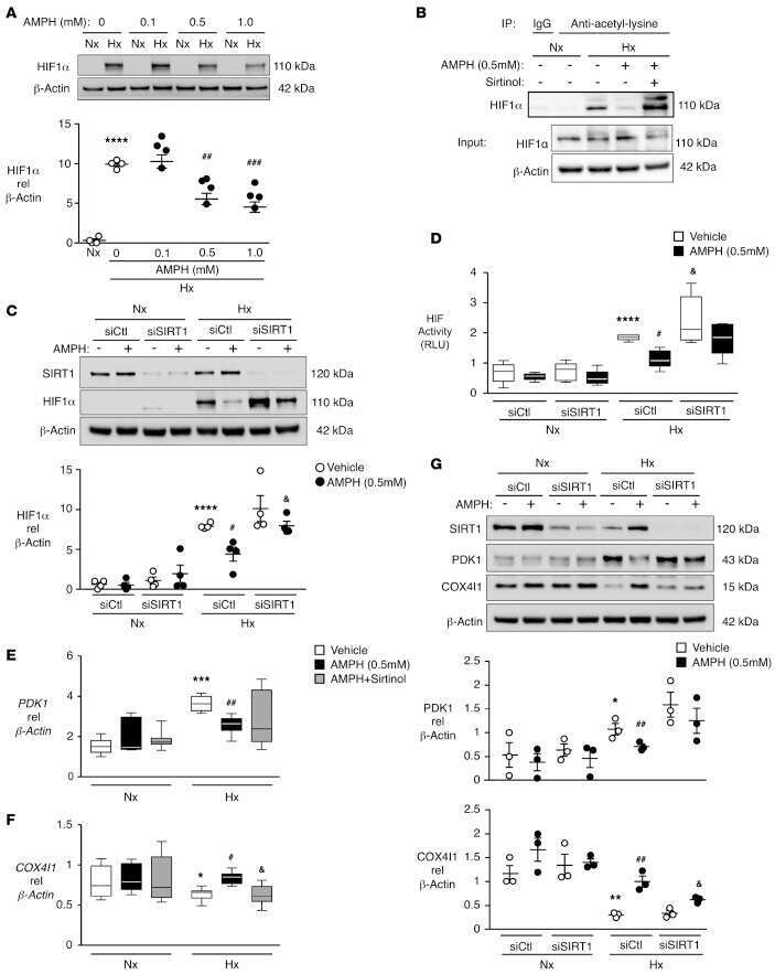
Amphetamines promote mitochondrial dysfunction and DNA damage in pulmonary hypertension.
Cartilage tissue formation using redifferentiated passaged chondrocytes in vitro.
Tammineni ER, Kraeva N, Figueroa L, Manno C, Ibarra CA, Klip A, Riazi S, Rios E
eLife 2020 May 4;9
eLife 2020 May 4;9
Isosteviol Protects Free Fatty Acid- and High Fat Diet-Induced Hepatic Injury via Modulating PKC-β/p66Shc/ROS and Endoplasmic Reticulum Stress Pathways.
Yi H, Xu D, Wu X, Xu F, Lin L, Zhou H
Antioxidants & redox signaling 2019 Jun 10;30(17):1949-1968
Antioxidants & redox signaling 2019 Jun 10;30(17):1949-1968
Amphetamines promote mitochondrial dysfunction and DNA damage in pulmonary hypertension.
Chen PI, Cao A, Miyagawa K, Tojais NF, Hennigs JK, Li CG, Sweeney NM, Inglis AS, Wang L, Li D, Ye M, Feldman BJ, Rabinovitch M
JCI insight 2017 Jan 26;2(2):e90427
JCI insight 2017 Jan 26;2(2):e90427
Cartilage tissue formation using redifferentiated passaged chondrocytes in vitro.
Ahmed N, Gan L, Nagy A, Zheng J, Wang C, Kandel RA
Tissue engineering. Part A 2009 Mar;15(3):665-73
Tissue engineering. Part A 2009 Mar;15(3):665-73
No comments: Submit comment
Supportive validation
- Submitted by
- Invitrogen Antibodies (provider)
- Main image
- Experimental details
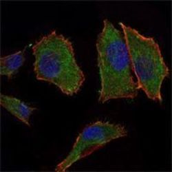
- Immunofluorescence analysis of PANC-1 cells using COX4I1 monoclonal antibody (Product # MA5-15686) (Green). Blue: DRAQ5 fluorescent DNA dye. Red: actin filaments have been labeled with phalloidin.

- Submitted by
- Invitrogen Antibodies (provider)
- Main image
- Experimental details
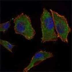
- Immunofluorescence analysis of PANC-1 cells using COX4I1 monoclonal antibody (Product # MA5-15686) (Green). Blue: DRAQ5 fluorescent DNA dye. Red: actin filaments have been labeled with phalloidin.
- Submitted by
- Invitrogen Antibodies (provider)
- Main image

- Experimental details
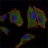
- Immunofluorescence analysis of PANC-1 cells using COX4I1 monoclonal antibody (Product # MA5-15686) (Green). Blue: DRAQ5 fluorescent DNA dye. Red: actin filaments have been labeled with phalloidin.

Supportive validation
- Submitted by
- Invitrogen Antibodies (provider)
- Main image
- Experimental details
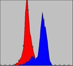
- Flow cytometric analysis of K562 cells using COX4I1 monoclonal antibody (Product # MA5-15686) (blue) and negative control (red).

Supportive validation
- Submitted by
- Invitrogen Antibodies (provider)
- Main image

- Experimental details
- NULL
- Submitted by
- Invitrogen Antibodies (provider)
- Main image
- Experimental details
- NULL

- Submitted by
- Invitrogen Antibodies (provider)
- Main image
- Experimental details
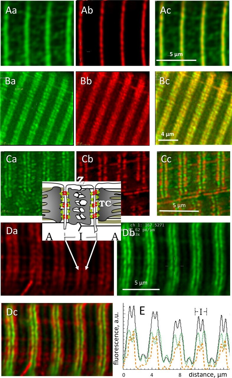
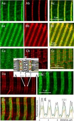
- Figure 4. Locating phosphorylated and apo forms of glycogen phosphorylase. Structure elements are diagrammed in inset. ( A ) Images of all-forms GP (Aa), Z disk component alpha-actinin (Ab) and their overlay (Ac) in a biopsy from an MHN subject (# 139). Shown are individual images ('sections') from separate emission ranges ('channels') in a 2-channel z -stack, after correction for blurring (Materials and methods). GP largely overlaps with actinin and is seen as low intensity patches at other locations. ( B ) All-forms GP (Ba), cytochrome c oxidase subunit 4 (Cox4), located in mitochondria (Bb) and overlay (Bc). Both proteins are located largely in the sarcomeric I band, in patches with limited overlap. ( C ) Myocyte from adult mouse FDB expressing GP-GFP (Ca), co-stained with mitochondrial marker TMRE (Cb). Most GP is in the I band, without actual overlap with TMRE (Cc). No GP is detected in the mitochondrial fraction extracted from these muscles ( Figure 4--figure supplement 1 ). ( D ) Human muscle cell co-stained for GP a and GP. Inset: location of Z disc and sarcomere bands I and A; TC, terminal cisternae of SR. GP a (Da) is distributed more broadly. Panel Dc shows in green a putative image of GP b (the apo form) obtained by subtracting from the all-forms GP fluorescence that of GP a , scaled by a factor that removes the 'shoulder' visible in the all-forms GP ( Equation 5 , Materials and methods). ( E ) The calculation is illustrated with image profiles F ( x ); black: F G