 Explore
 Validate
 Learn
Learn Western blot
Antibody data
- Antibody Data
- Antigen structure
- References [1]
- Comments [0]
- Validations
- Western blot [2]
- Immunocytochemistry [1]
- Immunohistochemistry [1]
Submit
Validation data
Reference
Comment
Report error
- Product number
- AF5814 - Provider product page

- Provider
- R&D Systems
- Product name
- Anti-Human/Mouse COX4-I1 Antigen Affinity-purified Polyclonal Antibody
- Antibody type
- Polyclonal
- Antigen
- E. coli-derived recombinant human COX4-I1, Ala23-Lys169
- Description
- Antigen Affinity-purified
- Reactivity
- Human, Mouse
- Host
- Goat
- Antigen sequence
P13073- Isotype
- IgG
- Vial size
- 100 µg
Submitted references Sarcolipin Signaling Promotes Mitochondrial Biogenesis and Oxidative Metabolism in Skeletal Muscle.
Maurya SK, Herrera JL, Sahoo SK, Reis FCG, Vega RB, Kelly DP, Periasamy M
Cell reports 2018 Sep 11;24(11):2919-2931
Cell reports 2018 Sep 11;24(11):2919-2931
No comments: Submit comment
Supportive validation
- Submitted by
- R&D Systems (provider)
- Main image
- Experimental details
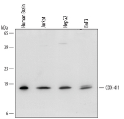
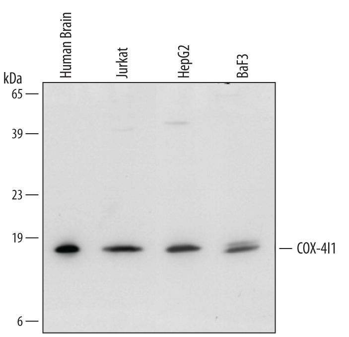
- Detection of Human/Mouse COX4-I1 by Western Blot. Western blot shows lysates of human brain tissue, Jurkat human acute T cell leukemia cell line, HepG2 human hepatocellular carcinoma cell line, and BaF3 mouse pro-B cell line. PVDF membrane was probed with 1 µg/mL of Goat Anti-Human/Mouse COX4-I1 Antigen Affinity-purified Polyclonal Antibody (Catalog # AF5814) followed by HRP-conjugated Anti-Goat IgG Secondary Antibody (Catalog # HAF109). A specific band was detected for COX4-I1 at approximately 18 kDa (as indicated). This experiment was conducted under reducing conditions and using Immunoblot Buffer Group 2.
- Submitted by
- R&D Systems (provider)
- Main image

- Experimental details
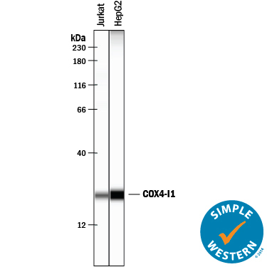
- Detection of Human COX4-I1 by Simple WesternTM. Simple Western lane view shows lysates of Jurkat human acute T cell leukemia cell line and HepG2 human hepatocellular carcinoma cell line, loaded at 0.2 mg/mL. A specific band was detected for COX4-I1 at approximately 24 kDa (as indicated) using 10 µg/mL of Goat Anti-Human/Mouse COX4-I1 Antigen Affinity-purified Polyclonal Antibody (Catalog # AF5814) followed by 1:50 dilution of HRP-conjugated Anti-Goat IgG Secondary Antibody (Catalog # HAF109). This experiment was conducted under reducing conditions and using the 12-230 kDa separation system.
Supportive validation
- Submitted by
- R&D Systems (provider)
- Main image
- Experimental details
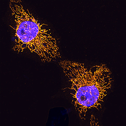
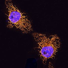
- COX4-I1 in HeLa Human Cell Line. COX4-I1 was detected in immersion fixed HeLa human cervical epithelial carcinoma cell line using Goat Anti-Human/Mouse COX4-I1 Antigen Affinity-purified Polyclonal Antibody (Catalog # AF5814) at 5 µg/mL for 3 hours at room temperature. Cells were stained using the NorthernLights™ 557-conjugated Anti-Goat IgG Secondary Antibody (yellow; Catalog # NL001) and counterstained with DAPI (blue). Specific staining was localized to mitochondria. View our protocol for Fluorescent ICC Staining of Cells on Coverslips.
Supportive validation
- Submitted by
- R&D Systems (provider)
- Main image
- Experimental details
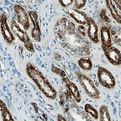
- COX4-I1 in Human Kidney. COX4-I1 was detected in immersion fixed paraffin-embedded sections of normal human kidney using Goat Anti-Human/Mouse COX4-I1 Antigen Affinity-purified Polyclonal Antibody (Catalog # AF5814) at 10 µg/mL overnight at 4 °C. Before incubation with the primary antibody, tissue was subjected to heat-induced epitope retrieval using Antigen Retrieval Reagent-Basic (Catalog # CTS013). Tissue was stained using the Anti-Goat HRP-DAB Cell & Tissue Staining Kit (brown; Catalog # CTS008) and counterstained with hematoxylin (blue). View our protocol for Chromogenic IHC Staining of Paraffin-embedded Tissue Sections.
