 Explore
 Validate
 Learn
Learn Western blot
Antibody data
- Antibody Data
- Antigen structure
- References [2]
- Comments [0]
- Validations
- Western blot [1]
- Immunocytochemistry [2]
Submit
Validation data
Reference
Comment
Report error
- Product number
- MAB6980 - Provider product page

- Provider
- R&D Systems
- Product name
- Human/Mouse COX4 Antibody
- Antibody type
- Monoclonal
- Description
- Protein A or G purified from hybridoma culture supernatant. Detects human COX4-I2 in direct ELISAs. In Western blots, 100% cross-reactivity with recombinant human (rh) COX4-I1 and no cross-reactivity with rhCOX-1 or rhCOX-2 is observed.
- Reactivity
- Human, Mouse
- Host
- Mouse
- Conjugate
- Unconjugated
- Antigen sequence
Q96KJ9- Isotype
- IgG
- Antibody clone number
- 673803
- Vial size
- 100 ug
- Concentration
- LYOPH
- Storage
- Use a manual defrost freezer and avoid repeated freeze-thaw cycles. 12 months from date of receipt, -20 to -70 °C as supplied. 1 month, 2 to 8 °C under sterile conditions after reconstitution. 6 months, -20 to -70 °C under sterile conditions after reconstitution.
Submitted references SIRT3 diminishes inflammation and mitigates endotoxin-induced acute lung injury.
Frataxin-deficient neurons and mice models of Friedreich ataxia are improved by TAT-MTScs-FXN treatment.
Kurundkar D, Kurundkar AR, Bone NB, Becker EJ Jr, Liu W, Chacko B, Darley-Usmar V, Zmijewski JW, Thannickal VJ
JCI insight 2019 Jan 10;4(1)
JCI insight 2019 Jan 10;4(1)
Frataxin-deficient neurons and mice models of Friedreich ataxia are improved by TAT-MTScs-FXN treatment.
Britti E, Delaspre F, Feldman A, Osborne M, Greif H, Tamarit J, Ros J
Journal of cellular and molecular medicine 2018 Feb;22(2):834-848
Journal of cellular and molecular medicine 2018 Feb;22(2):834-848
No comments: Submit comment
Supportive validation
- Submitted by
- R&D Systems (provider)
- Main image
- Experimental details
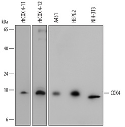
- Detection of Human and Mouse COX4 by Western Blot. Western blot shows lysates of A431 human epithelial carcinoma cell line, HepG2 human hepatocellular carcinoma cell line, and NIH-3T3 mouse embryonic fibroblast cell line. PVDF membrane was probed with 1 µg/mL of Mouse Anti-Human COX4 Monoclonal Antibody (Catalog # MAB6980) followed by HRP-conjugated Anti-Mouse IgG Secondary Antibody (Catalog # HAF007). For addtional reference, recombinant human COX4-I1 (5 ng/lane) and recombinant human COX4-I2 (2.5 ng/lane) were included. A specific band was detected for COX4 at approximately 18 kDa (as indicated). This experiment was conducted under reducing conditions and using Immunoblot Buffer Group 2.

Supportive validation
- Submitted by
- R&D Systems (provider)
- Main image
- Experimental details
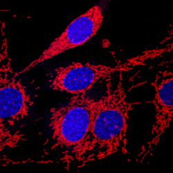
- COX4 in NIH-3T3 Mouse Cell Line. COX4 was detected in immersion fixed NIH-3T3 mouse embryonic fibroblast cell line using Mouse Anti-Human/Mouse COX4 Monoclonal Antibody (Catalog # MAB6980) at 10 µg/mL for 3 hours at room temperature. Cells were stained using the NorthernLights™ 557-conjugated Anti-Mouse IgG Secondary Antibody (red; Catalog # NL007) and counterstained with DAPI (blue). Specific staining was localized to mitochondria. View our protocol for Fluorescent ICC Staining of Cells on Coverslips.

- Submitted by
- R&D Systems (provider)
- Main image
- Experimental details
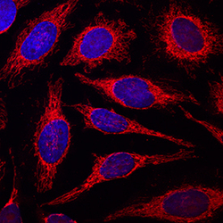
- COX4 in HeLa Human Cell Line. COX4 was detected in immersion fixed HeLa human cervical epithelial carcinoma cell line using Mouse Anti-Human/Mouse COX4 Monoclonal Antibody (Catalog # MAB6980) at 10 µg/mL for 3 hours at room temperature. Cells were stained using the NorthernLights™ 557-conjugated Anti-Mouse IgG Secondary Antibody (red; Catalog # NL007) and counterstained with DAPI (blue). Specific staining was localized to mitochondria. View our protocol for Fluorescent ICC Staining of Cells on Coverslips.
