Explore
 Validate
 Learn
Learn Western blot
 Immunocytochemistry
Antibody data
- Antibody Data
- Antigen structure
- References [2]
- Comments [0]
- Validations
- Immunocytochemistry [2]
- Immunohistochemistry [1]
- Other assay [1]
Submit
Validation data
Reference
Comment
Report error
- Product number
- PA5-82113 - Provider product page

- Provider
- Invitrogen Antibodies
- Product name
- OAS1 Polyclonal Antibody
- Antibody type
- Polyclonal
- Antigen
- Recombinant protein fragment
- Description
- Immunogen sequence: VFLSPLTTFQ DQLNRRGEFI QEIRRQLEAC QRERAFSVKF EVQAPRWGNP RALSFVLSSL QLGEGVEFDV LPAFDALGQL TGGYKPNPQI YVKLIEECTD LQKEGEFSTC FTELQRDFLK QRPTKLKSLI RLVKHWYQNC KK
- Reactivity
- Human
- Host
- Rabbit
- Isotype
- IgG
- Vial size
- 100 μL
- Concentration
- 0.2 mg/mL
- Storage
- Store at 4°C short term. For long term storage, store at -20°C, avoiding freeze/thaw cycles.
Submitted references Genetic regulation of OAS1 nonsense-mediated decay underlies association with COVID-19 hospitalization in patients of European and African ancestries.
Genetic regulation of OAS1 nonsense-mediated decay underlies association with risk of severe COVID-19.
Banday AR, Stanifer ML, Florez-Vargas O, Onabajo OO, Papenberg BW, Zahoor MA, Mirabello L, Ring TJ, Lee CH, Albert PS, Andreakos E, Arons E, Barsh G, Biesecker LG, Boyle DL, Brahier MS, Burnett-Hartman A, Carrington M, Chang E, Choe PG, Chisholm RL, Colli LM, Dalgard CL, Dude CM, Edberg J, Erdmann N, Feigelson HS, Fonseca BA, Firestein GS, Gehring AJ, Guo C, Ho M, Holland S, Hutchinson AA, Im H, Irby L, Ison MG, Joseph NT, Kim HB, Kreitman RJ, Korf BR, Lipkin SM, Mahgoub SM, Mohammed I, Paschoalini GL, Pacheco JA, Peluso MJ, Rader DJ, Redden DT, Ritchie MD, Rosenblum B, Ross ME, Anna HPS, Savage SA, Sharma S, Siouti E, Smith AK, Triantafyllia V, Vargas JM, Vargas JD, Verma A, Vij V, Wesemann DR, Yeager M, Yu X, Zhang Y, Boulant S, Chanock SJ, Feld JJ, Prokunina-Olsson L
Nature genetics 2022 Aug;54(8):1103-1116
Nature genetics 2022 Aug;54(8):1103-1116
Genetic regulation of OAS1 nonsense-mediated decay underlies association with risk of severe COVID-19.
Banday AR, Stanifer ML, Florez-Vargas O, Onabajo OO, Zahoor MA, Papenberg BW, Ring TJ, Lee CH, Andreakos E, Arons E, Barsh G, Biesecker LG, Boyle DL, Burnett-Hartman A, Carrington M, Chang E, Choe PG, Chrisholm RL, Dalgard C, Edberg J, Erdmann N, Feigelson HS, Firestein GS, Gehring AJ, Ho M, Holland S, Hutchinson AA, Im H, Ison MG, Kim HB, Kreitman RJ, Korf BR, Mirabello L, Pacheco JA, Peluso MJ, Rader DJ, Redden DT, Ritchie MD, Rosenbloom B, Sant Anna HP, Savage S, Siouti E, Triantafyllia V, Vargas JM, Verma A, Vij V, Wesemann DR, Yeager M, Yu X, Zhang Y, Boulant S, Chanock SJ, Feld JJ, Prokunina-Olsson L
medRxiv : the preprint server for health sciences 2021 Jul 13;
medRxiv : the preprint server for health sciences 2021 Jul 13;
No comments: Submit comment
Supportive validation
- Submitted by
- Invitrogen Antibodies (provider)
- Main image

- Experimental details
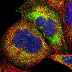
- Immunofluorescent analysis of OAS1 in A-431 cells using a OAS1 polyclonal antibody (Product # PA5-82113). The analysis shows localization to cytosol.

- Submitted by
- Invitrogen Antibodies (provider)
- Main image
- Experimental details
- Immunofluorecent analysis of OAS1 in human cell line A-431 using OAS1 Polyclonal Antibody (Product # PA5-82113). Staining shows localization to cytosol.

Supportive validation
- Submitted by
- Invitrogen Antibodies (provider)
- Main image
- Experimental details
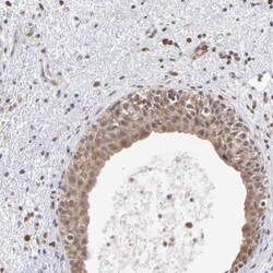
- Immunohistochemical analysis of OAS1 in human urinary bladder using OAS1 Polyclonal Antibody (Product # PA5-82113) shows cytoplasmic positivity in urothelial cells.

Supportive validation
- Submitted by
- Invitrogen Antibodies (provider)
- Main image
- Experimental details
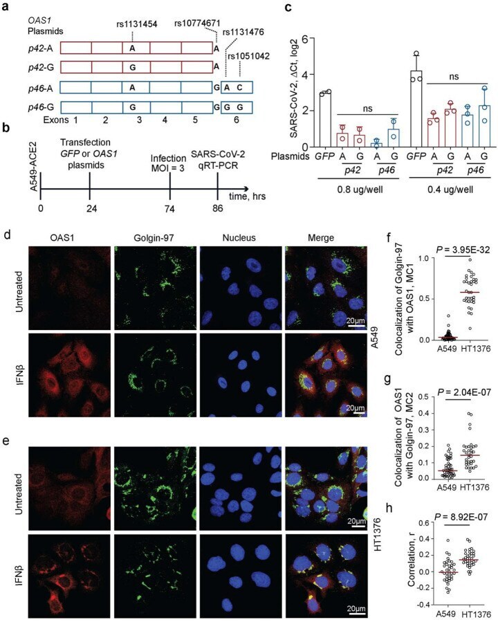
- Figure 2. Anti-SARS-CoV-2 activity and subcellular localization of OAS1-p42 and p46 isoforms. a) Description of OAS1 - p42 and OAS1 - p46 plasmids. b) Experiment outline: plasmids were transiently transfected in A549-ACE2 cells, followed by infection with SARS-CoV-2 and qRT-PCR for viral detection. c) SARS-CoV-2 load in A549-ACE2 cells transfected in duplicates or triplicates with 0.4 ug/well or 0.8 ug/well with OAS1 or GFP plasmids in six-well plates. Expression of SARS-CoV-2 was detected by qRT-PCR and normalized to the expression of an endogenous control ( HPRT1 ). P-values between OAS1 plasmids are for ANOVA tests. d, e) Representative confocal images for endogenous OAS1 expression in untreated and IFNbeta-treated A549 cells (rs10774671-AA, OAS1-p42, cytosolic expression) and HT1376 (rs10774671-GG, OAS1-p46, enrichment in trans-Golgi compartment); OAS1 (red), Golgin-97 (green) and nuclei (DAPI, blue). Scale bars, 20 mum. f) Mander's coefficient 1 (MC1) for colocalization of Golgin-97 with OAS1 in confocal images. g) Mander's coefficient 2 (MC2) for colocalization of OAS1 with Golgin-97 in confocal images. h) Overall correlation (Pearson's, r) between colocalization of Golgin-97 and OAS1 expression in confocal images. P -values are for non-parametric, two-sided Mann-Whitney U tests.