 Explore
 Validate
 Learn
Learn Western blot
Antibody data
- Antibody Data
- Antigen structure
- References [3]
- Comments [0]
- Validations
- Western blot [1]
- Immunohistochemistry [2]
Submit
Validation data
Reference
Comment
Report error
- Product number
- MAB1032-100 - Provider product page

- Provider
- R&D Systems
- Product name
- Human Cadherin-17 Antibody
- Antibody type
- Monoclonal
- Description
- Protein A or G purified from hybridoma culture supernatant. Detects human Cadherin-17 in direct ELISAs and Western blots. In direct ELISAs and Western blots, this antibody does not cross-react with recombinant human (rh) Cadherin-8, rhE-Cadherin, rhP-Cadherin, or rhVE-Cadherin.
- Reactivity
- Human
- Host
- Mouse
- Conjugate
- Unconjugated
- Antigen sequence
CAA58231- Isotype
- IgG
- Antibody clone number
- 141713
- Vial size
- 100 ug
- Concentration
- LYOPH
- Storage
- Use a manual defrost freezer and avoid repeated freeze-thaw cycles. 12 months from date of receipt, -20 to -70 °C as supplied. 1 month, 2 to 8 °C under sterile conditions after reconstitution. 6 months, -20 to -70 °C under sterile conditions after reconstitution.
Submitted references Expression Analysis of Fibronectin Type III Domain-Containing (FNDC) Genes in Inflammatory Bowel Disease and Colorectal Cancer.
Targeting cadherin-17 inactivates Ras/Raf/MEK/ERK signaling and inhibits cell proliferation in gastric cancer.
Differential protein expression on the cell surface of colorectal cancer cells associated to tumor metastasis.
Wuensch T, Wizenty J, Quint J, Spitz W, Bosma M, Becker O, Adler A, Veltzke-Schlieker W, Stockmann M, Weiss S, Biebl M, Pratschke J, Aigner F
Gastroenterology research and practice 2019;2019:3784172
Gastroenterology research and practice 2019;2019:3784172
Targeting cadherin-17 inactivates Ras/Raf/MEK/ERK signaling and inhibits cell proliferation in gastric cancer.
Lin Z, Zhang C, Zhang M, Xu D, Fang Y, Zhou Z, Chen X, Qin N, Zhang X
PloS one 2014;9(1):e85296
PloS one 2014;9(1):e85296
Differential protein expression on the cell surface of colorectal cancer cells associated to tumor metastasis.
Luque-García JL, Martínez-Torrecuadrada JL, Epifano C, Cañamero M, Babel I, Casal JI
Proteomics 2010 Mar;10(5):940-52
Proteomics 2010 Mar;10(5):940-52
No comments: Submit comment
Supportive validation
- Submitted by
- R&D Systems (provider)
- Main image
- Experimental details
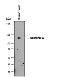
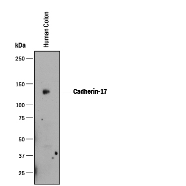
- Detection of Human Cadherin-17 by Western Blot. Western blot shows lysates of human colon tissue. PVDF membrane was probed with 1 µg/mL of Mouse Anti-Human Cadherin-17 Monoclonal Antibody (Catalog # MAB1032) followed by HRP-conjugated Anti-Mouse IgG Secondary Antibody (Catalog # HAF018). A specific band was detected for Cadherin-17 at approximately 125 kDa (as indicated). This experiment was conducted under reducing conditions and using Immunoblot Buffer Group 1.
Supportive validation
- Submitted by
- R&D Systems (provider)
- Main image
- Experimental details
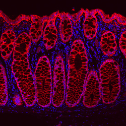
- Cadherin-17 in Human Transverse Colon. Cadherin-17 was detected in immersion fixed paraffin-embedded sections of human transverse colon using Mouse Anti-Human Cadherin-17 Monoclonal Antibody (Catalog # MAB1032) at 0.5 µg/mL overnight at 4 °C. Tissue was stained using the NorthernLights™ 557-conjugated Anti-Mouse IgG Secondary Antibody (red; Catalog # NL007) and counterstained with DAPI (blue).

- Submitted by
- R&D Systems (provider)
- Main image
- Experimental details
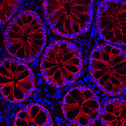
- Cadherin-17 in Human Ascending Colon. Cadherin-17 was detected in immersion fixed paraffin-embedded sections of human ascending colon using Mouse Anti-Human Cadherin-17 Monoclonal Antibody (Catalog # MAB1032) at 0.5 µg/mL overnight at 4 °C. Tissue was stained using the NorthernLights™ 557-conjugated Anti-Mouse IgG Secondary Antibody (red; Catalog # NL007) and counterstained with DAPI (blue).
