Explore
 Validate
 Learn
Learn Western blot
Antibody data
- Antibody Data
- Antigen structure
- References [0]
- Comments [0]
- Validations
- Western blot [2]
- Immunohistochemistry [1]
Submit
Validation data
Reference
Comment
Report error
- Product number
- MAB8524 - Provider product page

- Provider
- Novus Biologicals
- Product name
- Rabbit Monoclonal Cadherin-17 Antibody
- Antibody type
- Monoclonal
- Description
- Protein A or G purified from cell culture supernatant. Detects mouse Cadherin-17 in direct ELISAs and Western blots.
- Reactivity
- Mouse
- Host
- Rabbit
- Conjugate
- Unconjugated
- Isotype
- IgG
- Vial size
- 100 ug
- Storage
- Use a manual defrost freezer and avoid repeated freeze-thaw cycles. 12 months from date of receipt, -20 to -70 degreesC as supplied. 1 month, 2 to 8 degreesC under sterile conditions after reconstitution. 6 months, -20 to -70 degreesC under sterile conditions after reconstitution.
No comments: Submit comment
Supportive validation
- Submitted by
- Novus Biologicals (provider)
- Main image
- Experimental details
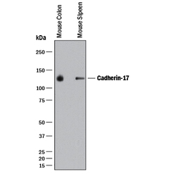
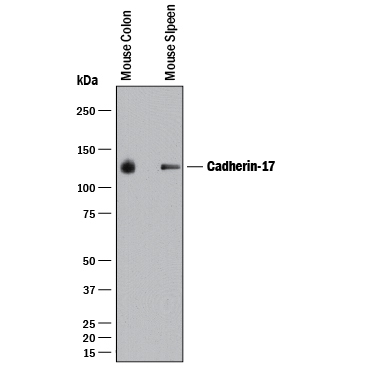
- Detection of Mouse Cadherin-17 by Western Blot. Western blot shows lysates of mouse colon tissue and mouse spleen tissue. PVDF membrane was probed with 0.1 µg/mL of Rabbit Anti-Mouse Cadherin-17 Monoclonal Antibody (Catalog # MAB8524) followed by HRP-conjugated Anti-Rabbit IgG Secondary Antibody (Catalog # HAF008). A specific band was detected for Cadherin-17 at approximately 130 kDa (as indicated). This experiment was conducted under reducing conditions and using Immunoblot Buffer Group 1.
- Submitted by
- Novus Biologicals (provider)
- Main image
- Experimental details
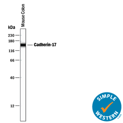
- Detection of Mouse Cadherin-17 by Simple WesternTM. Simple Western lane view shows lysates of mouse colon tissue, loaded at 0.2 mg/mL. A specific band was detected for Cadherin-17 at approximately 153 kDa (as indicated) using 1 µg/mL of Rabbit Anti-Mouse Cadherin-17 Monoclonal Antibody (Catalog # MAB8524) . This experiment was conducted under reducing conditions and using the 12-230 kDa separation system.

Supportive validation
- Submitted by
- Novus Biologicals (provider)
- Main image

- Experimental details
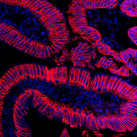
- Cadherin-17 in Mouse Intestine. Cadherin-17 was detected in perfusion fixed frozen sections of mouse intestine using Rabbit Anti-Mouse Cadherin-17 Monoclonal Antibody (Catalog # MAB8524) at 1.7 µg/mL overnight at 4 °C. Tissue was stained using the NorthernLights™ 557-conjugated Anti-Rabbit IgG Secondary Antibody (red; Catalog # NL004) and counterstained with DAPI (blue). Specific staining was localized to plasma membranes. View our protocol for Fluorescent IHC Staining of Frozen Tissue Sections.