Explore
 Validate
 Learn
Learn Western blot
 ELISA
ELISA Immunoprecipitation
Antibody data
- Antibody Data
- Antigen structure
- References [0]
- Comments [0]
- Validations
- Western blot [1]
- Immunocytochemistry [1]
Submit
Validation data
Reference
Comment
Report error
- Product number
- PA5-80458 - Provider product page

- Provider
- Invitrogen Antibodies
- Product name
- Anti-CDH17 Polyclonal Antibody
- Antibody type
- Polyclonal
- Antigen
- Recombinant full-length protein
- Description
- This product is preservative free. It is recommended to add sodium azide to avoid contamination (final concentration 0.05%-0.1%). This antibody has specificity for Human CDH17/Cadherin-17.
- Reactivity
- Human
- Host
- Rabbit
- Isotype
- IgG
- Vial size
- 100 µL
- Concentration
- Lot-Specific
- Storage
- Maintain refrigerated at 2-8°C for up to 1 month. For long term storage store at -20°C
No comments: Submit comment
Supportive validation
- Submitted by
- Invitrogen Antibodies (provider)
- Main image

- Experimental details
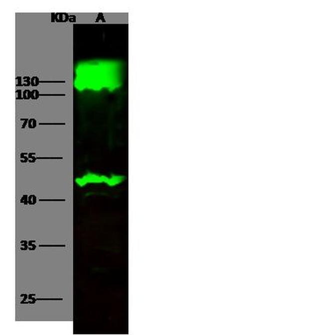
- Western blot analysis of CDH17 in Lane A: Jurkat Membrane Lysate (20 µg). Samples were probed using a CDH17 Polyclonal Antibody (Product # PA5-80458) at a 1:500 dilution, followed by a Goat Anti-Rabbit IgG (H+L), Dylight 800 Secondary Antibody at a 1:10000 dilution. Western blot was performed under reducing conditions. Predicted band size:92 kDa. Observed band size:80 kDa.
Supportive validation
- Submitted by
- Invitrogen Antibodies (provider)
- Main image
- Experimental details
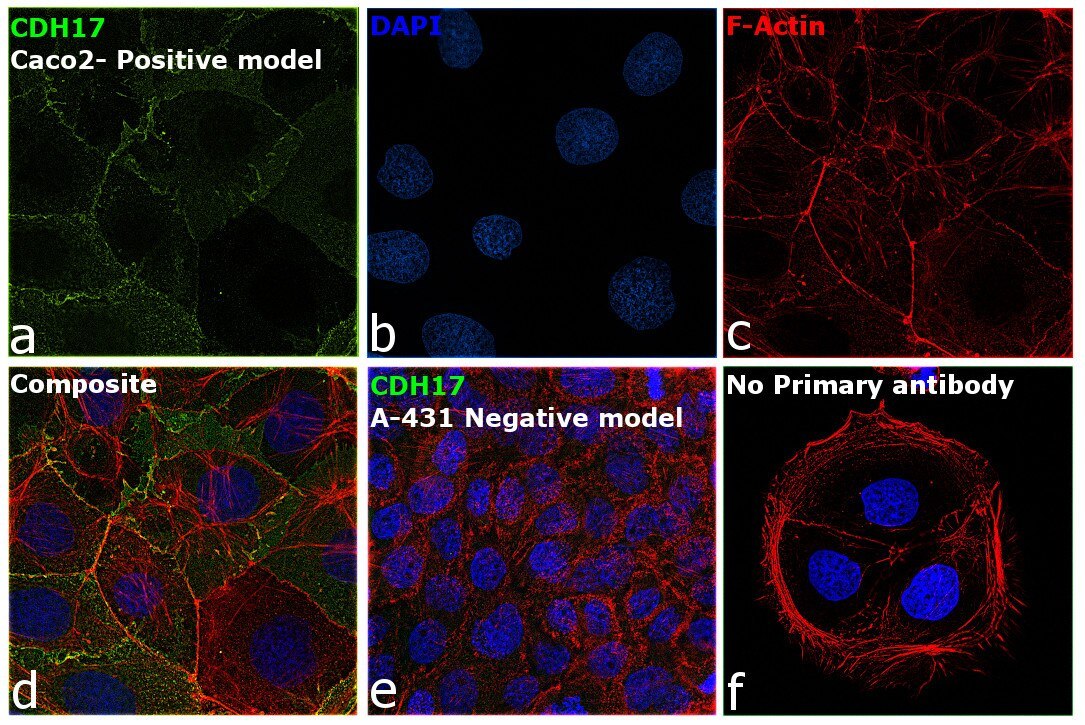
- Immunofluorescence analysis of Cadherin-17 was performed using 70% confluent log phase Caco-2 cells and A-431 cells. The cells were fixed with 4% paraformaldehyde for 10 minutes, permeabilized with 0.1% Triton™ X-100 for 15 minutes, and blocked with 2% BSA for 45 minutes at room temperature. The cells were labeled with CDH17 Polyclonal Antibody (Product # PA5-80458) at 1:100 in 0.1% BSA, incubated at 4 degree celsius overnight and then labeled with Donkey anti-Rabbit IgG (H+L) Highly Cross-Adsorbed Secondary Antibody, Alexa Fluor Plus 488 (Product # A32790), (1:2000 dilution), for 45 minutes at room temperature (Panel a: Green). Nuclei (Panel b: Blue) were stained with ProLong™ Diamond Antifade Mountant with DAPI (Product # P36962). F-actin (Panel c: Red) was stained with Rhodamine Phalloidin (Product # R415, 1:300). Panel d represents the merged image showing Plasma membrane localization in Caco-2 cells but not in A-431 cells (Panel e) which is reported to be low to negative for CDH17 expression. Panel f represents control cells with no primary antibody to assess background. The images were captured at 60X magnification.