Explore
 Validate
 Learn
Learn Western blot
 Immunocytochemistry
Antibody data
- Antibody Data
- Antigen structure
- References [1]
- Comments [0]
- Validations
- Immunocytochemistry [2]
- Immunohistochemistry [1]
- Other assay [2]
Submit
Validation data
Reference
Comment
Report error
- Product number
- PA5-54651 - Provider product page

- Provider
- Invitrogen Antibodies
- Product name
- RIPX Polyclonal Antibody
- Antibody type
- Polyclonal
- Antigen
- Recombinant protein fragment
- Description
- Immunogen sequence: MLEKDVCEKQ DALVSLRQQL DDLRALKHEL AFKLQSSDLG VKQKSELNSR LEEKTNQMAA TIKQLEQRLR QAERSRQSAE LDNRLFKQDF GDKINSLQLE VEELTRQRNQ LEL Highest antigen sequence identity to the following orthologs: Mouse - 97%, Rat - 97%.
- Reactivity
- Human, Mouse, Rat
- Host
- Rabbit
- Isotype
- IgG
- Vial size
- 100 μL
- Concentration
- 0.2 mg/mL
- Storage
- Store at 4°C short term. For long term storage, store at -20°C, avoiding freeze/thaw cycles.
Submitted references Roles of Rufy3 in experimental subarachnoid hemorrhage-induced early brain injury via accelerating neuronal axon repair and synaptic plasticity.
Wang Y, Xu J, You W, Shen H, Li X, Yu Z, Li H, Chen G
Molecular brain 2022 Apr 23;15(1):35
Molecular brain 2022 Apr 23;15(1):35
No comments: Submit comment
Supportive validation
- Submitted by
- Invitrogen Antibodies (provider)
- Main image

- Experimental details
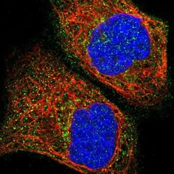
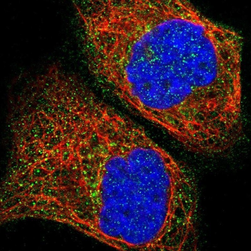
- Immunofluorescent staining of RIPX in human cell line A-431 using a RIPX Polyclonal Antibody (Product # PA5-54651) shows localization to cytosol.

- Submitted by
- Invitrogen Antibodies (provider)
- Main image
- Experimental details
- Immunofluorescent staining of RIPX in human cell line A-431 using a RIPX Polyclonal Antibody (Product # PA5-54651) shows localization to cytosol.
Supportive validation
- Submitted by
- Invitrogen Antibodies (provider)
- Main image
- Experimental details
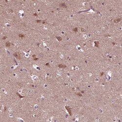
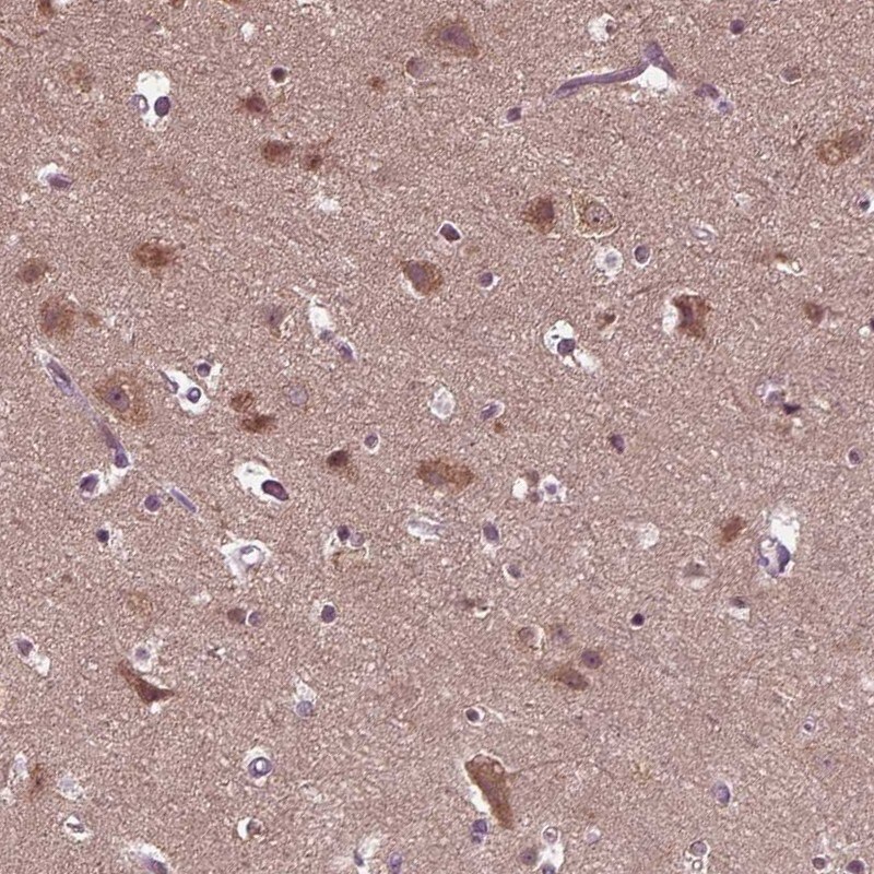
- Immunohistochemical staining of RIPX in human cerebral cortex using a RIPX Polyclonal Antibody (Product # PA5-54651) shows moderate cytoplasmic positivity in neuronal cells.
Supportive validation
- Submitted by
- Invitrogen Antibodies (provider)
- Main image
- Experimental details
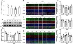
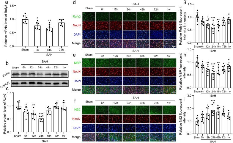
- Rufy3 mRNA and protein expression levels and neuronal axon damage following SAH. a Quantitative analysis of Rufy3 mRNA level. The sham group was used as a control. b Representative band of Rufy3 detected by western blot. c Quantitative analysis of Rufy3 at different stages following SAH. The sham group was used as a control. d - f Double immunofluorescence of Rufy3, MBP, and N52 (green, Alexa Fluor 488) and neuronal marker (NeuN; red, Alexa Fluor 555), and Rufy3 mainly located in the neurons. Nuclei were stained with DAPI (blue). Scale bars = 100 mum. g - i Immunopositivity of Rufy3, MBP, and N52 in neurons. The Sham group was used as the standard. Data are shown as mean +- SEM ( n = 6). * P < 0.05, ** P < 0.01, *** P < 0.01, vs. Sham group
- Submitted by
- Invitrogen Antibodies (provider)
- Main image
- Experimental details
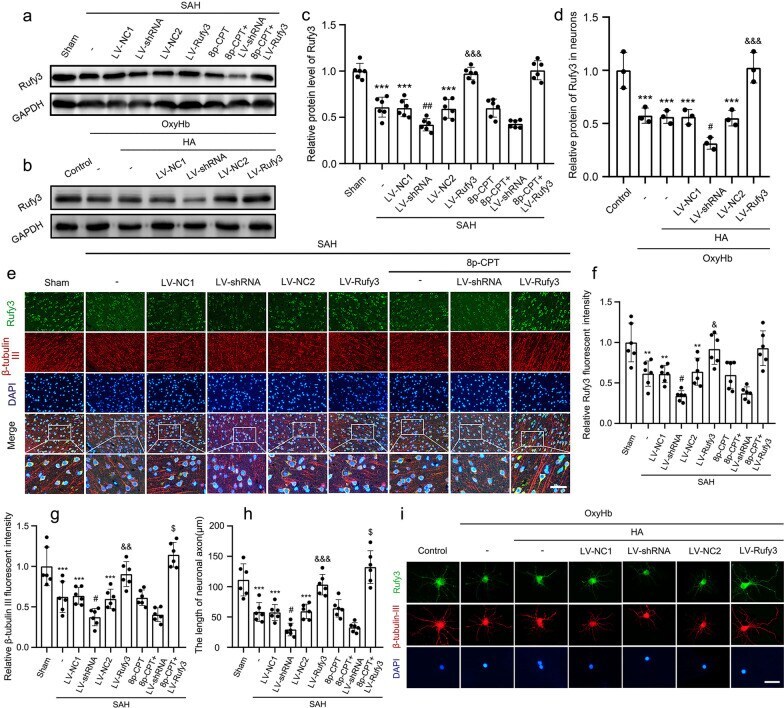
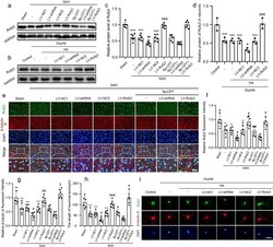
- The protein expression levels of Rufy3 and the state of neuronal axon under LV-shRNA and LV-Rufy3 treatments after vivo and vitro SAH. a Representative bands of Rufy3 detected by western blot under 8p-CPT, LV-shRNA and LV-Rufy3 treatments following vivo SAH. b Representative bands of Rufy3 detected by western blot under LV-shRNA and LV-Rufy3 treatments following vitro SAH. c , d Quantitative analysis of Rufy3 in different groups following vivo and vitro SAH. The sham and control group were used as a control. e Double immunofluorescence analysis of Rufy3 (green, Alexa Fluor 488) and beta-tubulin III (NeuN; red, Alexa Fluor 555); nuclei were stained with DAPI (blue). Scale bars = 32 mum. f, g Quantitative fluorescent intensity analysis of Rufy3 and beta-tubulin III expressions in different groups. The sham group was used as the standard. h Quantitative analysis of the length of neuronal axon in different groups. i Double immunofluorescence of Rufy3 (green, Alexa Fluor 488) and beta-tubulin III (axon; red, Alexa Fluor 555). Nuclei were stained with DAPI (blue). Scale bars = 100 mum. Data are shown as mean +- SEM ( n = 6). ** P < 0.01, ** P < 0.001 vs. Sham group; * P < 0.001 vs. Control group; # P < 0.05, ## P < 0.01 vs. LV-NC1 groups; & P < 0.05, && P < 0.01, &&& P < 0.001 vs. LV-NC2 group; $ P < 0.05 vs. LV-Rufy3 group