 Explore
 Validate
 Learn
Learn Western blot
 Immunocytochemistry
Antibody data
- Antibody Data
- Antigen structure
- References [0]
- Comments [0]
- Validations
- Western blot [1]
- Immunohistochemistry [1]
- Flow cytometry [1]
Submit
Validation data
Reference
Comment
Report error
- Product number
- MAB92171 - Provider product page

- Provider
- Novus Biologicals
- Product name
- Rabbit Monoclonal MCM7 Antibody
- Antibody type
- Monoclonal
- Description
- Protein A or G purified.
- Reactivity
- Human
- Host
- Rabbit
- Isotype
- IgG
- Vial size
- 100 ug
- Concentration
- 1.0 mg/ml
- Storage
- Store at -20C. Avoid freeze-thaw cycles.
No comments: Submit comment
Supportive validation
- Submitted by
- Novus Biologicals (provider)
- Main image

- Experimental details
- Western Blot: MCM7 Antibody (2068C) [MAB92171] - Analysis of HeLa, MCF-7, and MOLT-4, Daudi cell lysates with 0.5 ug/ml of hMCM7 Monoclonal Antibody followed by HRP-conjugated Anti-Rabbit IgG Antibody. A specific band was detected for hMCM7 at approximately 80 kDa (as indicated). This experiment was conducted under reducing conditions and using Immunoblot Buffer Group 1.

Supportive validation
- Submitted by
- Novus Biologicals (provider)
- Main image
- Experimental details
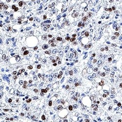
- Immunohistochemistry: MCM7 Antibody (2068C) [MAB92171] - MCM-7 was detected in immersion fixed paraffin-embedded sections of human mesothelioma using MCM-7 Monoclonal Antibody at 3 ug/ml. Tissue was stained using Rabbit IgG VisUCyte HRP Polymer Antibody (brown) and counterstained with hematoxylin (blue). Specific staining was localized to nuclei in cancer cells.

Supportive validation
- Submitted by
- Novus Biologicals (provider)
- Main image
- Experimental details
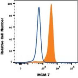
- Flow Cytometry: MCM7 Antibody (2068C) - Azide Free [MAB92171] - HeLa human cervical epithelial carcinoma cell line was stained with Rabbit Anti-Human MCM7 Monoclonal Antibody (Catalog # MAB92171, filled histogram) or isotype control antibody (Catalog # AB-105-C, open histogram), followed by Allophycocyanin-conjugated Anti-Rabbit IgG Secondary Antibody (Catalog # F0111). To facilitate intracellular staining, cells were fixed and permeabilized with FlowX FoxP3 Fixation & Permeabilization Buffer Kit (Catalog # FC012).
