Explore
 Validate
 Learn
Learn Western blot
 ELISA
ELISAAntibody data
- Antibody Data
- Antigen structure
- References [1]
- Comments [0]
- Validations
- Western blot [1]
- Immunocytochemistry [2]
- Other assay [1]
Submit
Validation data
Reference
Comment
Report error
- Product number
- PA5-98877 - Provider product page

- Provider
- Invitrogen Antibodies
- Product name
- EDG8 Polyclonal Antibody
- Antibody type
- Polyclonal
- Antigen
- Recombinant full-length protein
- Reactivity
- Human, Mouse
- Host
- Rabbit
- Isotype
- IgG
- Vial size
- 100 μg
- Concentration
- 1 mg/mL
- Storage
- -20°C or -80°C if preferred
Submitted references Inhibition of Sphingosine-1-Phosphate Receptor 2 by JTE013 Promoted Osteogenesis by Increasing Vesicle Trafficking, Wnt/Ca(2+), and BMP/Smad Signaling.
Lin S, Pandruvada S, Yu H
International journal of molecular sciences 2021 Nov 8;22(21)
International journal of molecular sciences 2021 Nov 8;22(21)
No comments: Submit comment
Supportive validation
- Submitted by
- Invitrogen Antibodies (provider)
- Main image
- Experimental details
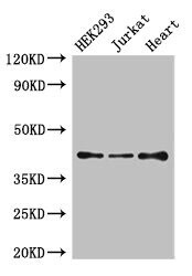
- Western Blot analysis of EDG8 using a EDG8 Polyclonal antibody (Product # PA5-98877) at a concentration of 4 µg/mL. Positive WB detected in: HEK293 whole cell lysate, Jurkat whole cell lysate, Mouse heart tissue. A secondary Goat polyclonal antibody to rabbit IgG was applied at a 1:50,000 dilution. Observed band size: 42 kDa.

Supportive validation
- Submitted by
- Invitrogen Antibodies (provider)
- Main image
- Experimental details
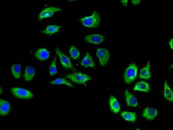
- Immunofluorescent analysis of EDG8 in A549 cells using a EDG8 polyclonal antibody (Product # PA5-98877) at a dilution of 1:100. Alexa Fluor 488-congugated Goat Anti-Rabbit IgG(H+L) secondary antibody was used.

- Submitted by
- Invitrogen Antibodies (provider)
- Main image

- Experimental details
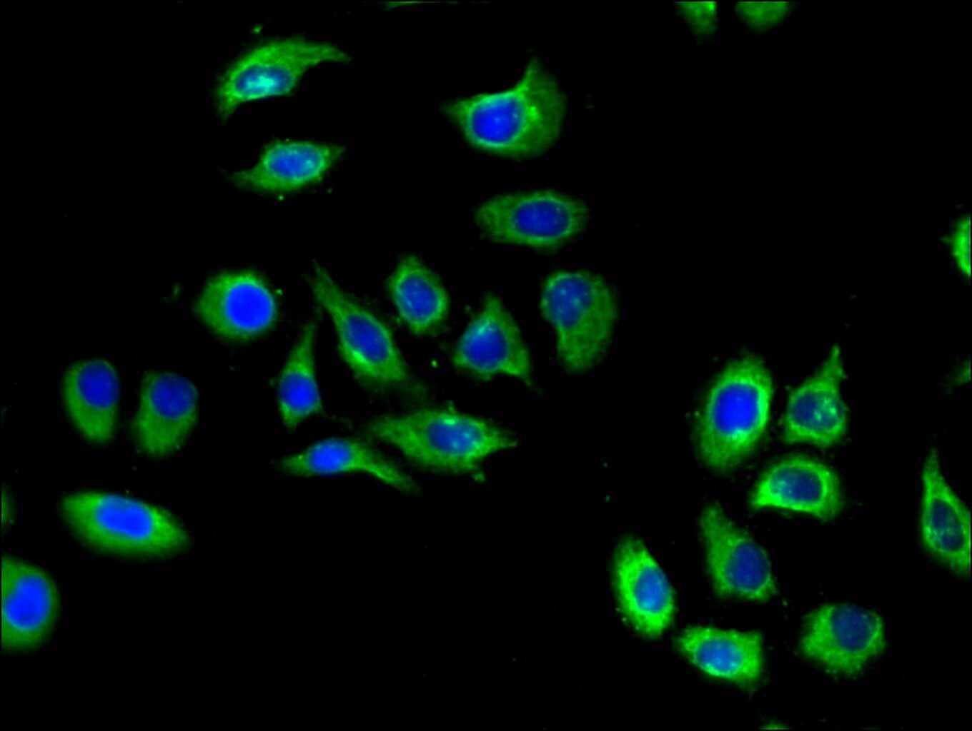
- Immunofluorescent analysis of EDG8 in A549 cells using a EDG8 polyclonal antibody (Product # PA5-98877) at a dilution of 1:100. Alexa Fluor 488-congugated Goat Anti-Rabbit IgG(H+L) secondary antibody was used.
Supportive validation
- Submitted by
- Invitrogen Antibodies (provider)
- Main image

- Experimental details
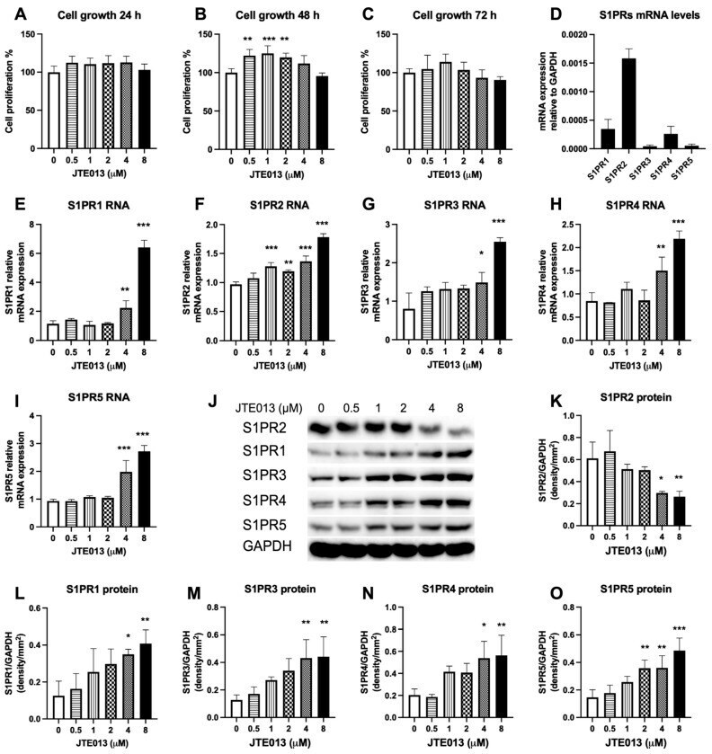
- Figure 1 Effects of JTE013 on cell growth and S1PRs expressions. Murine BMSCs were cultured in osteogenic media for 1 to 3 days with the presence of either DMSO or JTE013 (0.5 to 8 muM). Cell growth at ( A ) 24 h, ( B ) 48 h, and ( C ) 72 h was quantified by a CellTiter 96 (r) AQueous One Solution Cell Proliferation Assay. ( D ) Relative S1PRs' mRNA levels in murine BMSCs cultured in DMEM media with 10% FBS and 100 U/mL penicillin and streptomycin. ( E - I ) Murine BMSCs were treated with either DMSO or JTE013 (0.5 to 8 muM) and cultured in osteogenic media for 7 days. ( E ) S1PR1, ( F ) S1PR2, ( G ) S1PR3, ( H ) S1PR4, and ( I ) S1PR5 mRNA levels were quantified by RT-qPCR and normalized by GAPDH expression. (n = 3, * p < 0.05, ** p < 0.01, *** p < 0.001). ( J - O ) Murine BMSCs were treated with either DMSO or JTE013 (0.5 to 8 muM) and cultured in osteogenic media for 9 days. ( J ) S1PR2, S1PR1, S1PR3, S1PR4, S1PR5, and GAPDH protein levels were measured by Western blot. Protein densitometry of ( K ) S1PR2, ( L ) S1PR1, ( M ) S1PR3, ( N ) S1PR4, and ( O ) S1PR5 was normalized by GAPDH protein expression in BMSCs (n = 3, * p < 0.05, ** p < 0.01, *** p < 0.001).