Explore
 Validate
 Learn
Learn Western blot
 ELISA
ELISA Immunocytochemistry
Antibody data
- Antibody Data
- Antigen structure
- References [0]
- Comments [0]
- Validations
- Immunocytochemistry [6]
- Immunoprecipitation [1]
- Other assay [1]
Submit
Validation data
Reference
Comment
Report error
- Product number
- PA5-89181 - Provider product page

- Provider
- Invitrogen Antibodies
- Product name
- BAF57 Polyclonal Antibody
- Antibody type
- Polyclonal
- Antigen
- Recombinant full-length protein
- Description
- Immunogen sequence: MSKRPSYAPP PTPAPATQMP STPGFVGYNP YSHLAYNNYR LGGNPGTNSR VTASSGITIP KPPKPPDKPL MPYMRYSRKV WDQVKASNPD LKLWEIGKII GGMWRDLTDE EKQEYLNEYE AEKIEYNESM KAYHNSPAYL AYINAKSRAE AALEEESRQR QSRMEKGEPY MSIQPAEDPD DYDDGFSMKH TATARFQRNH RLISEILSES VVPDVRSVVT TARMQVLKRQ VQSLMVHQRK LEAELLQIEE RHQEKKRKFL ESTDSFNNEL KRLCGLKVEV DMEKIAAEIA QAEEQARKRQ EEREKEAAEQ AERSQSSIVP EEEQAANKGE EKKDDENIPM ETEETHLEET TESQQNGEEG TSTPEDKESG QEGVDSMAEE GTSDSNTGSE SNSATVEEPP TDPIPEDEKK E; Positive Samples: U-251MG, MCF-7; Cellular Location: Nucleus
- Reactivity
- Human, Mouse, Rat
- Host
- Rabbit
- Isotype
- IgG
- Vial size
- 100 μL
- Concentration
- 0.19 mg/mL
- Storage
- -20°C, Avoid Freeze/Thaw Cycles
No comments: Submit comment
Supportive validation
- Submitted by
- Invitrogen Antibodies (provider)
- Main image

- Experimental details
- Immunofluorescence analysis of HeLa cells using SMARCE1 Polyclonal antibody (Product # PA5-89181).

- Submitted by
- Invitrogen Antibodies (provider)
- Main image

- Experimental details
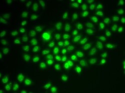
- Immunocytochemistry-Immunofluorescence analysis of BAF57 was performed in HeLa cells using BAF57 Polyclonal Antibody (Product # PA5-89181).

- Submitted by
- Invitrogen Antibodies (provider)
- Main image

- Experimental details
- Immunocytochemistry-Immunofluorescence analysis of BAF57 was performed in HeLa cells using BAF57 Polyclonal Antibody (Product # PA5-89181).

- Submitted by
- Invitrogen Antibodies (provider)
- Main image

- Experimental details
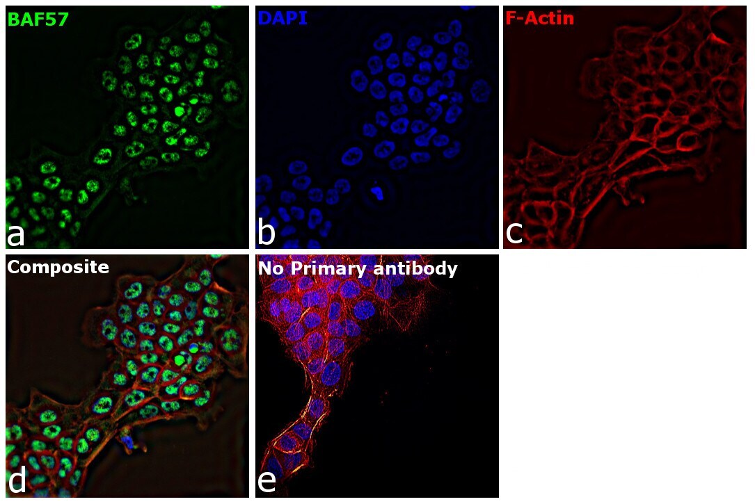
- Immunofluorescence analysis of BAF57 was performed using 70% confluent log phase A-431 cells. The cells were fixed with 4% paraformaldehyde for 10 minutes, permeabilized with 0.1% Triton™ X-100 for 15 minutes, and blocked with 2% BSA for 1 hour at room temperature. The cells were labeled with BAF57 Polyclonal Antibody (Product # PA5-89181) at 1:100 dilution in 0.1% BSA, incubated at 4 degree celsius overnight and then with Goat anti-Rabbit IgG (H+L) Highly Cross-Adsorbed Secondary Antibody, Alexa Fluor Plus 488 (Product # A32731) (1:2000) for 45 minutes at room temperature (Panel a: green). Nuclei (Panel b: blue) were stained with Hoechst 33342 (Product # H1399). F-actin (Panel c: red) was stained with Rhodamine Phalloidin (Product # R415, 1:300). Panel d represents the merged image showing nuclear localization. Panel e represents control cells with no primary antibody to assess background. The images were captured at 40X magnification in CellInsight CX7 LZR High-Content Screening (HCS) Platform (Product # CX7C1115LZR).
- Submitted by
- Invitrogen Antibodies (provider)
- Main image
- Experimental details
- Immunofluorescence analysis of BAF57 was performed using 70% confluent log phase A-431 cells. The cells were fixed with 4% paraformaldehyde for 10 minutes, permeabilized with 0.1% Triton™ X-100 for 15 minutes, and blocked with 2% BSA for 1 hour at room temperature. The cells were labeled with BAF57 Polyclonal Antibody (Product # PA5-89181) at 1:100 dilution in 0.1% BSA, incubated at 4 degree celsius overnight and then with Goat anti-Rabbit IgG (H+L) Highly Cross-Adsorbed Secondary Antibody, Alexa Fluor Plus 488 (Product # A32731) (1:2000) for 45 minutes at room temperature (Panel a: green). Nuclei (Panel b: blue) were stained with Hoechst 33342 (Product # H1399). F-actin (Panel c: red) was stained with Rhodamine Phalloidin (Product # R415, 1:300). Panel d represents the merged image showing nuclear localization. Panel e represents control cells with no primary antibody to assess background. The images were captured at 40X magnification in CellInsight CX7 LZR High-Content Screening (HCS) Platform (Product # CX7C1115LZR).

- Submitted by
- Invitrogen Antibodies (provider)
- Main image
- Experimental details
- Immunofluorescence analysis of BAF57 in HeLa cells. Samples were incubated with BAF57 Polyclonal antibody (Product # PA5-89181).

Supportive validation
- Submitted by
- Invitrogen Antibodies (provider)
- Main image

- Experimental details
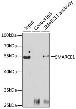
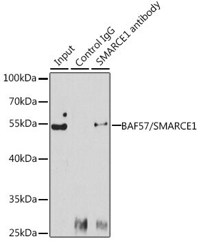
- Immunoprecipitation of BAF57 in 150 μg extracts of Jurkat cells. Samples were precipitated with 3 μg BAF57 Polyclonal antibody (Product # PA5-89181). Western blot was performed from the immunoprecipitate using BAF57 Polyclonal antibody (Product # PA5-89181) at a dilution of 1:1,000.
Supportive validation
- Submitted by
- Invitrogen Antibodies (provider)
- Main image

- Experimental details
- Immunoprecipitation analysis of BAF57 was performed in 150 µg extracts of Jurkat cells using BAF57 Polyclonal Antibody (Product # PA5-89181). Western blot was performed from the immunoprecipitate using BAF57 Polyclonal Antibody.