Explore
 Validate
 Learn
LearnMAB943-100
antibody from R&D Systems
Targeting: TNFRSF25
APO-3, DDR3, DR3, LARD, TNFRSF12, TR3, TRAMP, WSL-1, WSL-LR
 Western blot
Antibody data
- Antibody Data
- Antigen structure
- References [1]
- Comments [0]
- Validations
- Western blot [1]
- Flow cytometry [2]
Submit
Validation data
Reference
Comment
Report error
- Product number
- MAB943-100 - Provider product page

- Provider
- R&D Systems
- Product name
- Human/Mouse DR3/TNFRSF25 Antibody
- Antibody type
- Monoclonal
- Description
- Protein A or G purified from ascites. Detects human DR3/TNFRSF25 in direct ELISAs and Western blots. Shows 100% cross-reactivity with recombinant mouse (rm) DR3/TNFRSF25 and no cross-reactivity with recombinant human (rh) 4-1BB, rhCD27, rhCD30, rhCD40, rhDR6, rhBAFF R, rhFas, rhGITR, rhLTR beta , rhNGF R, rhOPG, rmOX40, rhRANK, rhTAJ, rhTNF RI, rhTNF RII, or rhHVEM.
- Reactivity
- Human, Mouse
- Host
- Mouse
- Conjugate
- Unconjugated
- Antigen sequence
Q93038- Isotype
- IgG
- Antibody clone number
- 59204
- Vial size
- 100 ug
- Storage
- Use a manual defrost freezer and avoid repeated freeze-thaw cycles. 12 months from date of receipt, -20 to -70 °C as supplied. 1 month, 2 to 8 °C under sterile conditions after reconstitution. 6 months, -20 to -70 °C under sterile conditions after reconstitution.
Submitted references Survival advantages conferred to colon cancer cells by E-selectin-induced activation of the PI3K-NFκB survival axis downstream of Death receptor-3.
Porquet N, Poirier A, Houle F, Pin AL, Gout S, Tremblay PL, Paquet ER, Klinck R, Auger FA, Huot J
BMC cancer 2011 Jul 1;11:285
BMC cancer 2011 Jul 1;11:285
No comments: Submit comment
Supportive validation
- Submitted by
- R&D Systems (provider)
- Main image

- Experimental details
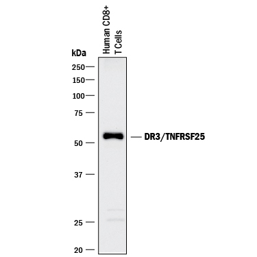
- Detection of Human DR3/TNFRSF25 by Western Blot. Western blot shows lysates of human CD8+ T cells. PVDF membrane was probed with 2 µg/mL of Mouse Anti-Human/Mouse DR3/TNFRSF25 Monoclonal Antibody (Catalog # MAB943) followed by HRP-conjugated Anti-Mouse IgG Secondary Antibody (Catalog # HAF018). A specific band was detected for DR3/TNFRSF25 at approximately 55 kDa (as indicated). This experiment was conducted under reducing conditions and using Immunoblot Buffer Group 1.
Supportive validation
- Submitted by
- R&D Systems (provider)
- Main image

- Experimental details
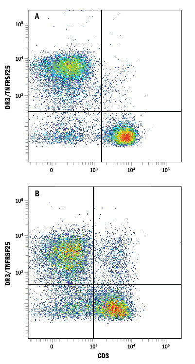
- Detection of DR3/TNFRSF25 in Human PBMCs by Flow Cytometry. Human peripheral blood mononuclear cells (PBMCs) either (A) untreated or (B) treated with PMA and Calcium Ionomycin for 24 hours were stained with Mouse Anti-Human DR3/TNFRSF25 Monoclonal Antibody (Catalog # MAB943) followed by Phycoerythrin-conjugated Anti-Mouse IgG Secondary Antibody (Catalog # F0102B) and Mouse Anti-Human CD3 epsilon APC-conjugated Monoclonal Antibody (Catalog # FAB100A). Quadrant markers were set based on control antibody staining (Catalog # MAB002).
- Submitted by
- R&D Systems (provider)
- Main image
- Experimental details
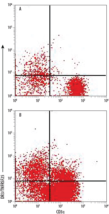
- Detection of DR3/TNFRSF25 in Mouse Splenocytes by Flow Cytometry. Mouse splenocytes either (A) untreated or (B) treated with PMA and Calcium Ionomycin for 24 hours were stained with Mouse Anti-Human DR3/TNFRSF25 Monoclonal Antibody (Catalog # MAB943) followed by Allophycocyanin-conjugated Anti-Mouse IgG Secondary Antibody (Catalog # F0101B) and Rat Anti-Mouse CD3 PE-conjugated Monoclonal Antibody (Catalog # FAB4841P). Quadrant markers were set based on control antibody staining (Catalog # MAB002).