Explore
 Validate
 Learn
Learn Western blot
 Immunocytochemistry
Antibody data
- Antibody Data
- Antigen structure
- References [6]
- Comments [0]
- Validations
- Immunocytochemistry [2]
- Immunohistochemistry [1]
- Flow cytometry [2]
- Other assay [4]
Submit
Validation data
Reference
Comment
Report error
- Product number
- PA5-23696 - Provider product page

- Provider
- Invitrogen Antibodies
- Product name
- FUS Polyclonal Antibody
- Antibody type
- Polyclonal
- Antigen
- Synthetic peptide
- Description
- This antibody is predicted to react with bovine based on sequence homology.
- Reactivity
- Human, Mouse
- Host
- Rabbit
- Isotype
- IgG
- Vial size
- 400 μL
- Concentration
- 0.5 mg/mL
- Storage
- Store at 4°C short term. For long term storage, store at -20°C, avoiding freeze/thaw cycles.
Submitted references Muscle cells of sporadic amyotrophic lateral sclerosis patients secrete neurotoxic vesicles.
FUS-induced circular RNA ZNF609 promotes tumorigenesis and progression via sponging miR-142-3p in lung cancer.
Extensive splicing changes in an ALS/FTD transgenic mouse model overexpressing cytoplasmic fused in sarcoma.
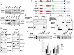
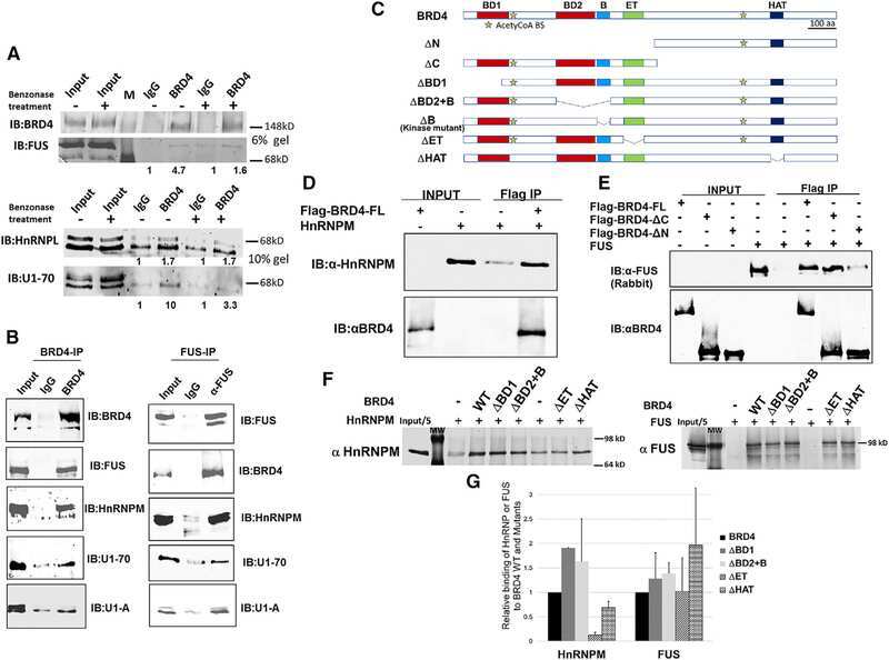
The Bromodomain Protein 4 Contributes to the Regulation of Alternative Splicing.
Dendritic Homeostasis Disruption in a Novel Frontotemporal Dementia Mouse Model Expressing Cytoplasmic Fused in Sarcoma.
Mislocated FUS is sufficient for gain-of-toxic-function amyotrophic lateral sclerosis phenotypes in mice.
Le Gall L, Duddy WJ, Martinat C, Mariot V, Connolly O, Milla V, Anakor E, Ouandaogo ZG, Millecamps S, Lainé J, Vijayakumar UG, Knoblach S, Raoul C, Lucas O, Loeffler JP, Bede P, Behin A, Blasco H, Bruneteau G, Del Mar Amador M, Devos D, Henriques A, Hesters A, Lacomblez L, Laforet P, Langlet T, Leblanc P, Le Forestier N, Maisonobe T, Meininger V, Robelin L, Salachas F, Stojkovic T, Querin G, Dumonceaux J, Butler Browne G, González De Aguilar JL, Duguez S, Pradat PF
Journal of cachexia, sarcopenia and muscle 2022 Apr;13(2):1385-1402
Journal of cachexia, sarcopenia and muscle 2022 Apr;13(2):1385-1402
FUS-induced circular RNA ZNF609 promotes tumorigenesis and progression via sponging miR-142-3p in lung cancer.
Liu S, Yang N, Jiang X, Wang J, Dong J, Gao Y
Journal of cellular physiology 2021 Jan;236(1):79-92
Journal of cellular physiology 2021 Jan;236(1):79-92
Extensive splicing changes in an ALS/FTD transgenic mouse model overexpressing cytoplasmic fused in sarcoma.
Ito D, Taguchi R, Deguchi M, Ogasawara H, Inoue E
Scientific reports 2020 Mar 17;10(1):4857
Scientific reports 2020 Mar 17;10(1):4857
The Bromodomain Protein 4 Contributes to the Regulation of Alternative Splicing.
Uppal S, Gegonne A, Chen Q, Thompson PS, Cheng D, Mu J, Meerzaman D, Misra HS, Singer DS
Cell reports 2019 Nov 19;29(8):2450-2460.e5
Cell reports 2019 Nov 19;29(8):2450-2460.e5
Dendritic Homeostasis Disruption in a Novel Frontotemporal Dementia Mouse Model Expressing Cytoplasmic Fused in Sarcoma.
Shiihashi G, Ito D, Arai I, Kobayashi Y, Hayashi K, Otsuka S, Nakajima K, Yuzaki M, Itohara S, Suzuki N
EBioMedicine 2017 Oct;24:102-115
EBioMedicine 2017 Oct;24:102-115
Mislocated FUS is sufficient for gain-of-toxic-function amyotrophic lateral sclerosis phenotypes in mice.
Shiihashi G, Ito D, Yagi T, Nihei Y, Ebine T, Suzuki N
Brain : a journal of neurology 2016 Sep;139(Pt 9):2380-94
Brain : a journal of neurology 2016 Sep;139(Pt 9):2380-94
No comments: Submit comment
Supportive validation
- Submitted by
- Invitrogen Antibodies (provider)
- Main image

- Experimental details
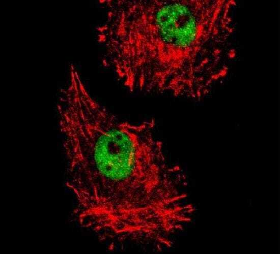
- Immunofluorescent analysis of MDA-MB231 cells using a FUS polyclonal antibody (Product # PA5-23696) at a dilution of 1:10-50. Primary antibody was detected with goat anti-rabbit lgG, fluor-conjugated secondary antibody (green). Actin filaments have been labeled with red dye conjugated phalloidin.
- Submitted by
- Invitrogen Antibodies (provider)
- Main image
- Experimental details
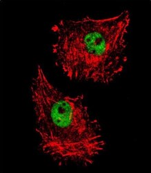
- Immunocytochemistry analysis of FUS in MDA-MB231 cells. Samples were incubated in FUS polyclonal antibody (Product # PA5-23696) followed by Alexa Fluor 488-conjugated goat anti-rabbit lgG (green). Actin filaments have been labeled with Alexa Fluor 555 phalloidin (red).
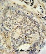
Supportive validation
- Submitted by
- Invitrogen Antibodies (provider)
- Main image
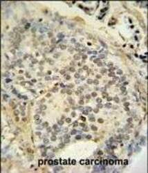
- Experimental details
- Immunohistochemistry analysis of FUS in formalin fixed and paraffin embedded human prostate carcinoma. Samples were incubated with FUS polyclonal antibody (Product # PA5-23696) followed by peroxidase conjugation of the secondary antibody and DAB staining. This data demonstrates the use of this antibody for immunohistochemistry. Clinical relevance has not been evaluated.
Supportive validation
- Submitted by
- Invitrogen Antibodies (provider)
- Main image
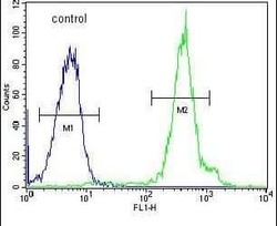
- Experimental details
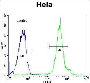
- Flow cytometry analysis of HeLa cells using a FUS polyclonal antibody (Product # PA5-23696) (right) compared to a negative control cell (left) at a dilution of 1:10-50, followed by a FITC-conjugated goat anti-rabbit antibody
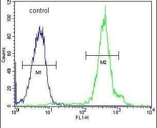
- Submitted by
- Invitrogen Antibodies (provider)
- Main image

- Experimental details
- Flow cytometry of FUS in Hela cells (right histogram). Samples were incubated with FUS polyclonal antibody (Product # PA5-23696) followed by FITC-conjugated goat-anti-rabbit secondary antibody. Negative control cell (left histogram).
Supportive validation
- Submitted by
- Invitrogen Antibodies (provider)
- Main image
- Experimental details
- NULL
- Submitted by
- Invitrogen Antibodies (provider)
- Main image
- Experimental details
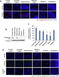
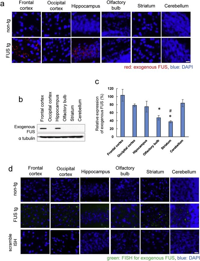
- Fig. 1 Expression of exogenous FUS in transgenic mice at 15 weeks of age. (a) Immunohistochemistry with anti-Myc (red) antibody in each part of the brain. Sections counterstained with 4',6-diamidino-2-phenylindole (DAPI; blue). Scale bar, 10 mum. (b) Western blot analysis of brain lysates using anti-Myc antibody. Expression of FUS with nuclear localization signal deletion (DeltaNLS-FUS) protein appears restricted to the frontal cortex and hippocampus. alpha-tubulin was used as an internal control. (c) Quantitative reverse transcriptase polymerase chain reaction (qRT-PCR) analysis for exogenous FUS mRNA in the brain of tg mice. ( n = 3 per genotype; * P < 0.05 vs. frontal, occipital cortex and cerebellum; #P < 0.05 vs. hippocampus by Student's t -test). (d) In situ hybridization showing the expression of the exogenous FUS mRNA (green) in the brain of tg mice. Scale bar, 10 mum. Fig. 1
- Submitted by
- Invitrogen Antibodies (provider)
- Main image
- Experimental details
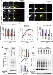
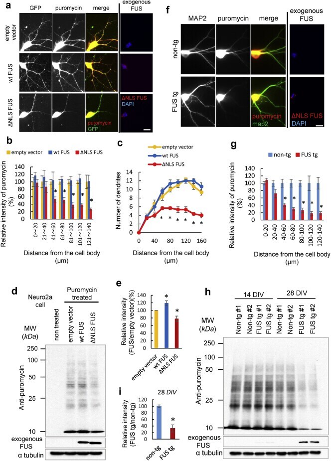
- Fig. 7 Fused in sarcoma (FUS) with nuclear localization signal deletion (DeltaNLS-FUS) but not wild-type FUS impaired protein synthesis. (a) Protein synthesis was detected by anti-puromycin antibody (red) in the primary cultured neurons transfected with GFP and empty vector, wild-type FUS, or DeltaNLS-FUS after pulse-treatment with puromycin. Dendrites were identified by GFP expression. Scale bar, 20 mum. (b) Quantification of dendritic protein synthesis as assessed by puromycylation in Fig. 7 a. The fluorescence intensities of each group were normalized to the mean GFP signal in each dendritic segment. The graphs show the percentage of the mean puromycin signal (mean +- standard error) in each transfected cell to the empty vector group in each dendritic segment. Fifteen transfected cells from three independent experiments were analyzed. (c) Quantitative data on the number of dendritic branches in the primary cultured neurons expressing wild-type FUS or DeltaNLS-FUS. n = 15 per group. (d) Neuro2a cells were transfected with empty vector, wild-type FUS, or DeltaNLS-FUS, and then treated with puromycin. Cell lysates were analyzed by western blotting using an anti-puromycin antibody. (e) Quantification of dendritic protein synthesis as assessed by puromycylation in Fig. 7 d. The graphs show the intensities of puromycylation in each cell relative to that of the empty vector (mean +- standard error; n = 3). *P < 0.05 vs. empty vector by Student's t -test. (f) Primary cultured
- Submitted by
- Invitrogen Antibodies (provider)
- Main image
- Experimental details
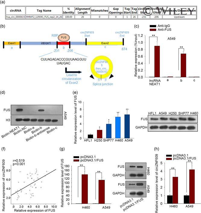
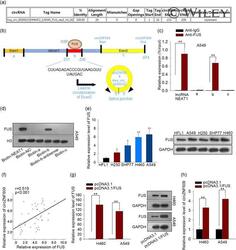
- 3 Figure Upregulation of circZNF609 is associated with FUS RNA-binding protein. (a) CircInteractome predicted that FUS bounded to upstream -31 to -206 of circZNF609 pre-mRNA. (b) Schematic representation of the binding sites of FUS in the upstream region of circZNF609 pre-mRNA. The binding region was labeled as b, exon1 was labeled as a, and exon2 was labeled as c. (c) RIP assay indicated that anti-FUS can immunoprecipitate b region. LncRNA- NEAT1 reported to be combinative to FUS was positive control. Anti-IgG was used as negative control. (d) RNA pull-down suggested that biotinylated b region could pulldown FUS. (e) mRNA and protein levels of FUS in LC cells and normal cells were measured. (f) Expression correlation between circZNF609 and FUS in LC tissues was analyzed by the Pearson correlation test. (g) qPCR and Western blotting examined the overexpression efficiency of FUS. (h) qRT-PCR demonstrated that the ectopic expression of FUS significantly promoted the circZNF609 expression. All experiments were conducted in triplicate. * p < .05, ** p < .01. LC, lung cancer; mRNA, messenger RNA; qRT-PCR, quantitative real-time polymerase chain reaction; RIP, RNA-binding protein immunoprecipitation