Explore
 Validate
 Learn
Learn Western blot
Antibody data
- Antibody Data
- Antigen structure
- References [10]
- Comments [0]
- Validations
- Western blot [3]
- Immunoprecipitation [2]
- Immunohistochemistry [5]
Submit
Validation data
Reference
Comment
Report error
- Product number
- NB100-561 - Provider product page

- Provider
- Novus Biologicals
- Proper citation
- Novus Cat#NB100-561, RRID:AB_10002260
- Product name
- Rabbit Polyclonal FUS Antibody
- Antibody type
- Polyclonal
Submitted references Cytoplasmic Restriction of Mutated SOD1 Impairs the DNA Repair Process in Spinal Cord Neurons.
Human proteins that interact with RNA/DNA hybrids.
Position-dependent FUS-RNA interactions regulate alternative splicing events and transcriptions.
Localization of fused in sarcoma (FUS) protein to the post-synaptic density in the brain.
Dysfunction of the ubiquitin-proteasome system in the cerebellum of aging Ts65Dn mice.
Mutant FUS proteins that cause amyotrophic lateral sclerosis incorporate into stress granules.
FUS mutations in familial amyotrophic lateral sclerosis in the Netherlands.
TDRD3, a novel Tudor domain-containing protein, localizes to cytoplasmic stress granules.
The Ewing sarcoma protein (EWS) binds directly to the proximal elements of the macrophage-specific promoter of the CSF-1 receptor (csf1r) gene.
The Ewing sarcoma protein (EWS) binds directly to the proximal elements of the macrophage-specific promoter of the CSF-1 receptor (csf1r) gene.
Li J, Song M, Moh S, Kim H, Kim DH
Cells 2019 Nov 23;8(12)
Cells 2019 Nov 23;8(12)
Human proteins that interact with RNA/DNA hybrids.
Wang IX, Grunseich C, Fox J, Burdick J, Zhu Z, Ravazian N, Hafner M, Cheung VG
Genome research 2018 Sep;28(9):1405-1414
Genome research 2018 Sep;28(9):1405-1414
Position-dependent FUS-RNA interactions regulate alternative splicing events and transcriptions.
Ishigaki S, Masuda A, Fujioka Y, Iguchi Y, Katsuno M, Shibata A, Urano F, Sobue G, Ohno K
Scientific reports 2012;2:529
Scientific reports 2012;2:529
Localization of fused in sarcoma (FUS) protein to the post-synaptic density in the brain.
Aoki N, Higashi S, Kawakami I, Kobayashi Z, Hosokawa M, Katsuse O, Togo T, Hirayasu Y, Akiyama H
Acta neuropathologica 2012 Sep;124(3):383-94
Acta neuropathologica 2012 Sep;124(3):383-94
Dysfunction of the ubiquitin-proteasome system in the cerebellum of aging Ts65Dn mice.
Necchi D, Lomoio S, Scherini E
Experimental neurology 2011 Dec;232(2):114-8
Experimental neurology 2011 Dec;232(2):114-8
Mutant FUS proteins that cause amyotrophic lateral sclerosis incorporate into stress granules.
Bosco DA, Lemay N, Ko HK, Zhou H, Burke C, Kwiatkowski TJ Jr, Sapp P, McKenna-Yasek D, Brown RH Jr, Hayward LJ
Human molecular genetics 2010 Nov 1;19(21):4160-75
Human molecular genetics 2010 Nov 1;19(21):4160-75
FUS mutations in familial amyotrophic lateral sclerosis in the Netherlands.
Groen EJ, van Es MA, van Vught PW, Spliet WG, van Engelen-Lee J, de Visser M, Wokke JH, Schelhaas HJ, Ophoff RA, Fumoto K, Pasterkamp RJ, Dooijes D, Cuppen E, Veldink JH, van den Berg LH
Archives of neurology 2010 Feb;67(2):224-30
Archives of neurology 2010 Feb;67(2):224-30
TDRD3, a novel Tudor domain-containing protein, localizes to cytoplasmic stress granules.
Goulet I, Boisvenue S, Mokas S, Mazroui R, Côté J
Human molecular genetics 2008 Oct 1;17(19):3055-74
Human molecular genetics 2008 Oct 1;17(19):3055-74
The Ewing sarcoma protein (EWS) binds directly to the proximal elements of the macrophage-specific promoter of the CSF-1 receptor (csf1r) gene.
Hume DA, Sasmono T, Himes SR, Sharma SM, Bronisz A, Constantin M, Ostrowski MC, Ross IL
Journal of immunology (Baltimore, Md. : 1950) 2008 May 15;180(10):6733-42
Journal of immunology (Baltimore, Md. : 1950) 2008 May 15;180(10):6733-42
No comments: Submit comment
Supportive validation
- Submitted by
- Novus Biologicals (provider)
- Main image
- Experimental details
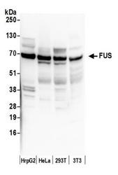
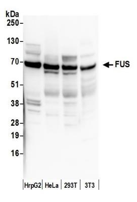
- Western Blot: FUS Antibody [NB100-561] - Detection of Human and Mouse FUS by Western Blot. Samples: Whole cell lysate (50 ug) from HepG2, HeLa, 293T, and mouse NIH3T3 cells prepared using NETN lysis buffer. Antibody: Affinity purified rabbit anti-FUS antibody NB100-561 used for WB at 0.1 ug/ml. Detection: Chemiluminescence with an exposure time of 30 seconds.
- Submitted by
- Novus Biologicals (provider)
- Main image

- Experimental details
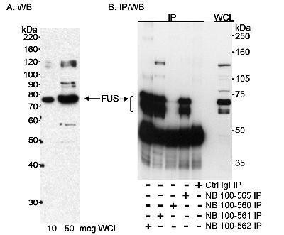
- Western Blot: FUS Antibody [NB100-561] - Detection of human FUS on HeLa whole cell lysate using NB100-561. For IP in B, rabbit anti-FUS antibodies NB100-560, NB100-561, and NB100-565 were used.
- Submitted by
- Novus Biologicals (provider)
- Main image
- Experimental details
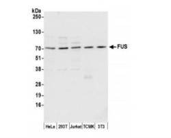
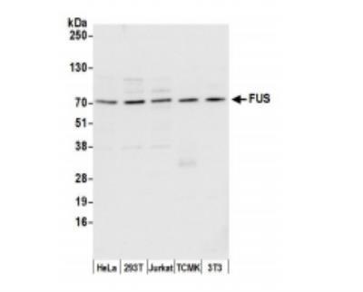
- Western Blot: FUS Antibody [NB100-561] - Whole cell lysate (15 ug) from HeLa, HEK293T, Jurkat, mouse TCMK-1, and mouse NIH 3T3 cells prepared using NETN lysis buffer. Antibody: Affinity purified rabbit anti-FUS antibody used for WB at 0.1 ug/ml. Detection: Chemiluminescence with an exposure time of 3 seconds.
Supportive validation
- Submitted by
- Novus Biologicals (provider)
- Main image

- Experimental details
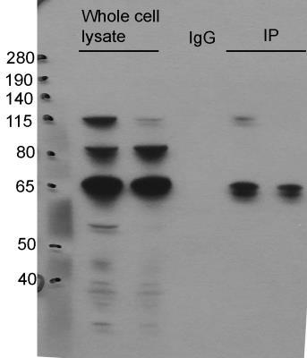
- Immunoprecipitation: FUS Antibody [NB100-561] - FUS immunoprecipitation in human fibroblast cell lysates. Image from verified customer review.
- Submitted by
- Novus Biologicals (provider)
- Main image
- Experimental details
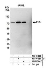
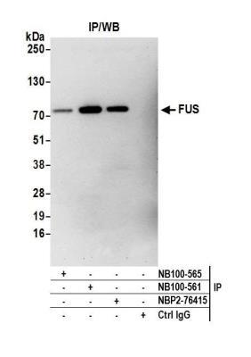
- Immunoprecipitation: FUS Antibody [NB100-561] - Detection of human FUS by western blot of immunoprecipitates. Samples: Whole cell lysate (1.0 mg per IP reaction; 20% of IP loaded) from HEK293T cells prepared using NETN lysis buffer. Antibodies: Affinity purified rabbit anti-FUS antibody NB100-561 used for IP at 3 ug per reaction. FUS was also immunoprecipitated by rabbit anti-FUS recombinant monoclonal antibody [BLR023E] (NBP2-76415) and rabbit anti-FUS antibody NB100-565. For blotting immunoprecipitated FUS, NBP2-76415 was used at 1:1000. Detection: Chemiluminescence with an exposure time time of 3 minutes.
Supportive validation
- Submitted by
- Novus Biologicals (provider)
- Main image
- Experimental details
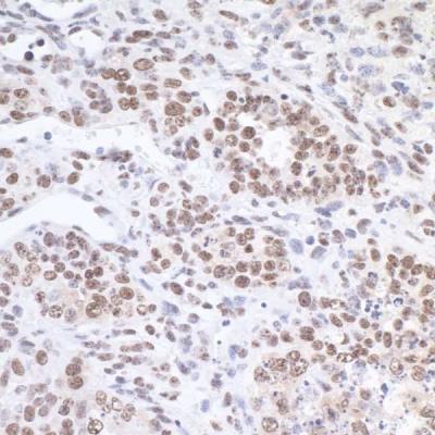
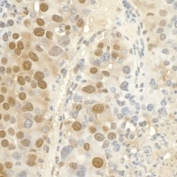
- Immunohistochemistry: FUS Antibody [NB100-561] - Sample: FFPE section of mouse renal cell carcinoma. Antibody: Affinity purified rabbit anti-FUS used at a dilution of 1:1,000 (1ug/ml). Detection: DAB
- Submitted by
- Novus Biologicals (provider)
- Main image
- Experimental details
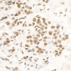
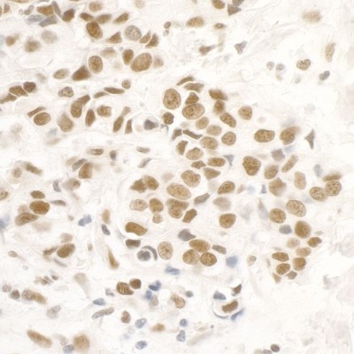
- Immunohistochemistry: FUS Antibody [NB100-561] - Sample: FFPE section of human breast carcinoma. Antibody: Affinity purified rabbit anti-FUS used at a dilution of 1:5,000 (0.2ug/ml). Detection: DAB
- Submitted by
- Novus Biologicals (provider)
- Main image

- Experimental details
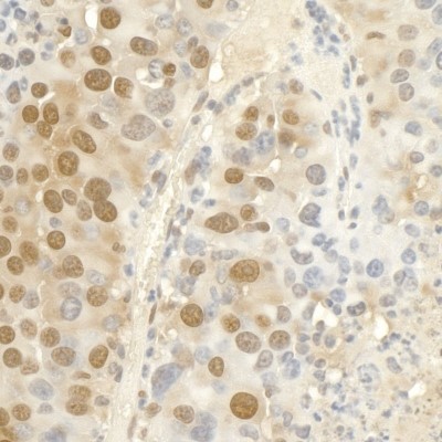
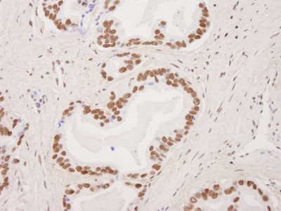
- Immunohistochemistry-Paraffin: FUS Antibody [NB100-561] - Human prostate carcinoma. Antibody used at a dilution of 1:1,000.
- Submitted by
- Novus Biologicals (provider)
- Main image

- Experimental details
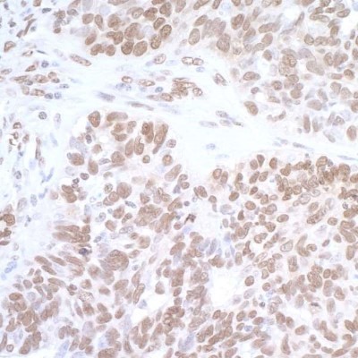
- Immunohistochemistry-Paraffin: FUS Antibody [NB100-561] - Section of human ovarian carcinoma. Antibody: Affinity purified rabbit anti-FUS (NB100-561). Detection: DAB
- Submitted by
- Novus Biologicals (provider)
- Main image
- Experimental details
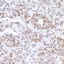
- Immunohistochemistry-Paraffin: FUS Antibody [NB100-561] - Detection of mouse FUS by immunohistochemistry. Sample: FFPE section of mouse teratoma Antibody: Affinity purified rabbit anti-FUS (NB100-561) used at 1:5,000 (0.2ug/ml). Detection: DAB