 Explore
 Validate
 Learn
Learn Western blot
Antibody data
- Antibody Data
- Antigen structure
- References [0]
- Comments [0]
- Validations
- Western blot [1]
- Immunocytochemistry [1]
- Immunohistochemistry [1]
- Flow cytometry [1]
Submit
Validation data
Reference
Comment
Report error
- Product number
- AP51735PU-N - Provider product page

- Provider
- Acris Antibodies GmbH
- Proper citation
- Acris Antibodies GmbH Cat#AP51735PU-N, RRID:AB_11149296
- Product name
- anti FUS / TLS (C-term)
- Antibody type
- Polyclonal
- Antigen
- KLH conjugated synthetic peptide between 506-534 amino acids from the C-terminal region of human FUS
- Reactivity
- Human, Mouse
- Host
- Rabbit
- Vial size
- 0.4 ml
- Concentration
- lot specific
No comments: Submit comment
Supportive validation
- Submitted by
- Acris Antibodies GmbH (provider)
- Main image
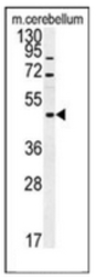
- Experimental details
- Western blot analysis of FUS Antibody (C-term) Cat.-No AP51735PU-NÂ in mouse cerebellum tissue lysates (15ug/lane).This demonstrates the FUS antibody detected FUS protein (arrow).

Supportive validation
- Submitted by
- Acris Antibodies GmbH (provider)
- Main image
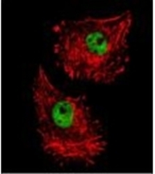
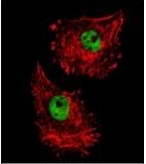
- Experimental details
- Confocal immunofluorescent analysis of FUS Antibody (C-term) Cat.-No AP51735PU-N with MDA-MB231 cell followed by Alexa Fluor 488-conjugated goat anti-rabbit lgG (green).Actin filaments have been labeled with Alexa Fluor 555 phalloidin (red).
Supportive validation
- Submitted by
- Acris Antibodies GmbH (provider)
- Main image
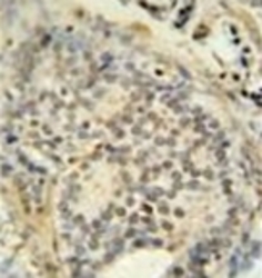
- Experimental details
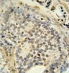
- Immunohistochemistry analysis in formalin fixed and paraffin embedded human prostate carcinoma stained with FUS Antibody (C-term) Cat.-No AP51735PU-Nfollowed by peroxidase conjugation of the secondary antibody and DAB staining. This data demonstrates the use of the FUS antibody (C-term) for immunohistochemistry. Clinical relevance has not been evaluated.
Supportive validation
- Submitted by
- Acris Antibodies GmbH (provider)
- Main image
- Experimental details
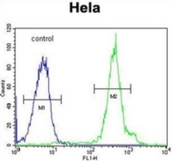
- Flow cytometric analysis of Hela cells using FUS Antibody (C-term) Cat.-No AP51735PU-N (right histogram) compared to a negative control cell (left histogram).FITC-conjugated goat-anti-rabbit secondary antibodies were used for the analysis.
