Explore
 Validate
 Learn
Learn Western blot
 Immunoprecipitation
Antibody data
- Antibody Data
- Antigen structure
- References [3]
- Comments [0]
- Validations
- Western blot [3]
- Immunocytochemistry [3]
- Immunohistochemistry [1]
Submit
Validation data
Reference
Comment
Report error
- Product number
- GTX115154 - Provider product page

- Provider
- GeneTex
- Proper citation
- GeneTex Cat#GTX115154, RRID:AB_10729580
- Product name
- FUBP1 antibody
- Antibody type
- Polyclonal
- Reactivity
- Human, Mouse, Rat
- Host
- Rabbit
Submitted references Additive Promotion of Viral Internal Ribosome Entry Site-Mediated Translation by Far Upstream Element-Binding Protein 1 and an Enterovirus 71-Induced Cleavage Product.
Resources for the Comprehensive Discovery of Functional RNA Elements.
Nuclear proteomics with XRCC3 knockdown to reveal the development of doxorubicin-resistant uterine cancer.
Hung CT, Kung YA, Li ML, Brewer G, Lee KM, Liu ST, Shih SR
PLoS pathogens 2016 Oct;12(10):e1005959
PLoS pathogens 2016 Oct;12(10):e1005959
Resources for the Comprehensive Discovery of Functional RNA Elements.
Sundararaman B, Zhan L, Blue SM, Stanton R, Elkins K, Olson S, Wei X, Van Nostrand EL, Pratt GA, Huelga SC, Smalec BM, Wang X, Hong EL, Davidson JM, Lécuyer E, Graveley BR, Yeo GW
Molecular cell 2016 Mar 17;61(6):903-13
Molecular cell 2016 Mar 17;61(6):903-13
Nuclear proteomics with XRCC3 knockdown to reveal the development of doxorubicin-resistant uterine cancer.
Chang JF, Lin ST, Hung E, Lu YL, Soon May EW, Lo YW, Chou HC, Chan HL
Toxicological sciences : an official journal of the Society of Toxicology 2014 Jun;139(2):396-406
Toxicological sciences : an official journal of the Society of Toxicology 2014 Jun;139(2):396-406
No comments: Submit comment
Supportive validation
- Submitted by
- GeneTex (provider)
- Main image
- Experimental details
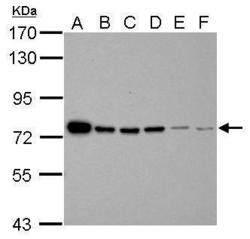
- Sample (30 ug of whole cell lysate) A: Jurkat B: Raji C: K562 D: THP-1 E: HL-60 F: NCI-H929 7.5% SDS PAGE GTX115154 diluted at 1:5000

- Submitted by
- GeneTex (provider)
- Main image
- Experimental details
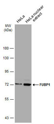
- Hela whole cell and nuclear extracts (30 ?g) were separated by 7.5% SDS-PAGE, and the membrane was blotted with FUBP1 antibody (GTX115154) diluted at 1:5000. The HRP-conjugated anti-rabbit IgG antibody (GTX213110-01) was used to detect the primary antibody.
- Submitted by
- GeneTex (provider)
- Main image
- Experimental details
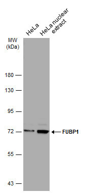
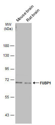
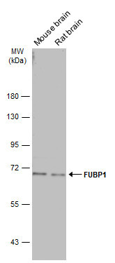
- Various tissue extracts (50 ?g) were separated by 7.5% SDS-PAGE, and the membrane was blotted with FUBP1 antibody (GTX115154) diluted at 1:5000. The HRP-conjugated anti-rabbit IgG antibody (GTX213110-01) was used to detect the primary antibody.
Supportive validation
- Submitted by
- GeneTex (provider)
- Main image
- Experimental details
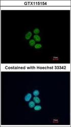
- Immunofluorescence analysis of paraformaldehyde-fixed MCF-7, using FUBP1(GTX115154) antibody at 1:500 dilution.

- Submitted by
- GeneTex (provider)
- Main image

- Experimental details
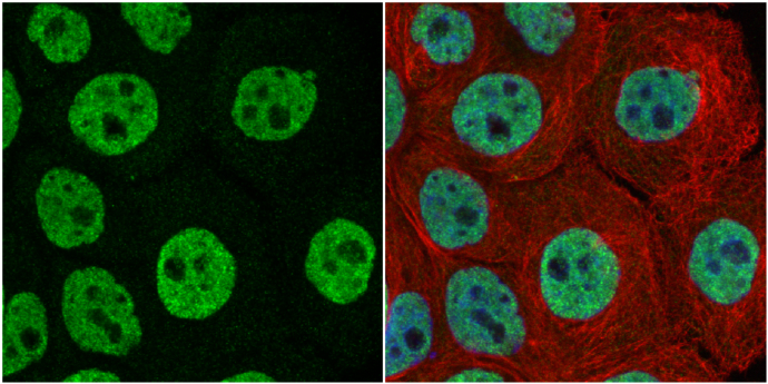
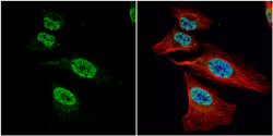
- FUBP1 antibody detects FUBP1 protein at nucleus by immunofluorescent analysis.Sample: A431 cells were fixed in 4% paraformaldehyde at RT for 15 min.Green: FUBP1 protein stained by FUBP1 antibody (GTX115154) diluted at 1:1000.Red: alpha Tubulin, a cytoskeleton marker, stained by alpha Tubulin antibody [GT114] (GTX628802) diluted at 1:1000.Blue: Hoechst 33342 staining.
- Submitted by
- GeneTex (provider)
- Main image
- Experimental details
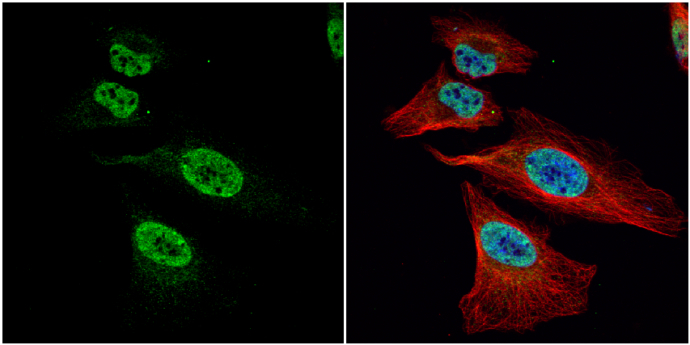
- FUBP1 antibody detects FUBP1 protein at nucleus by immunofluorescent analysis.Sample: HeLa cells were fixed in 4% paraformaldehyde at RT for 15 min.Green: FUBP1 protein stained by FUBP1 antibody (GTX115154) diluted at 1:1000.Red: alpha Tubulin, a cytoskeleton marker, stained by alpha Tubulin antibody [B-5-1-2] (GTX11304) diluted at 1:10000.Blue: Hoechst 33342 staining.
Supportive validation
- Submitted by
- GeneTex (provider)
- Main image
- Experimental details
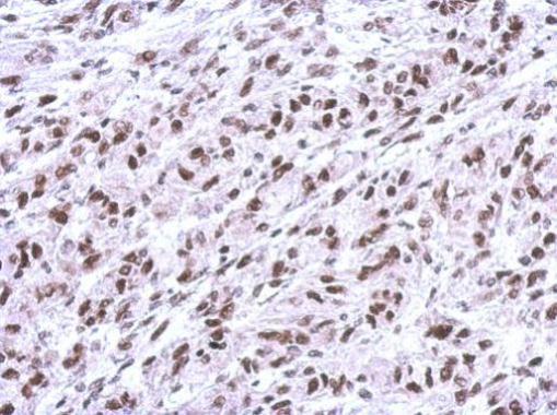
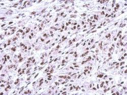
- Immunohistochemical analysis of paraffin-embedded U373 xenograft, using FUBP1(GTX115154) antibody at 1:500 dilution.