Explore
 Validate
 Learn
Learn Western blot
 Immunocytochemistry
Antibody data
- Antibody Data
- Antigen structure
- References [0]
- Comments [0]
- Validations
- Immunocytochemistry [2]
- Immunoprecipitation [1]
- Immunohistochemistry [2]
- Flow cytometry [2]
- Other assay [1]
Submit
Validation data
Reference
Comment
Report error
- Product number
- MA5-38293 - Provider product page

- Provider
- Invitrogen Antibodies
- Product name
- FUBP1 Recombinant Rabbit Monoclonal Antibody (7C3)
- Antibody type
- Monoclonal
- Antigen
- Synthetic peptide
- Description
- This antibody has been tested in direct-ELISA
- Reactivity
- Human
- Host
- Rabbit
- Isotype
- IgG
- Antibody clone number
- 7C3
- Vial size
- 100 μL
- Concentration
- 0.39 mg/mL
- Storage
- -20°C or -80°C if preferred
No comments: Submit comment
Supportive validation
- Submitted by
- Invitrogen Antibodies (provider)
- Main image
- Experimental details
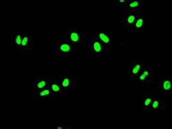
- Immunocytochemistry/Immunofluorescence analysis of FUBP1 in HeLa cells using FUBP1 Monoclonal Antibody (Product # MA5-38293) diluted at 1:50, counter-stained with DAPI. The cells were fixed in 4% formaldehyde, permeated by 0.2% TritonX-100, and blocked in 10% normal Goat Serum. The cells were then incubated with the antibody overnight at 4°C. Nuclear DNA was labeled in blue with DAPI. The secondary antibody was FITC-conjugated AffiniPure Goat Anti-Rabbit IgG (H+L).

- Submitted by
- Invitrogen Antibodies (provider)
- Main image

- Experimental details
- Immunocytochemistry/Immunofluorescence analysis of FUBP1 in HeLa cells using FUBP1 Monoclonal Antibody (Product # MA5-38293) diluted at 1:50, counter-stained with DAPI. The cells were fixed in 4% formaldehyde, permeated by 0.2% TritonX-100, and blocked in 10% normal Goat Serum. The cells were then incubated with the antibody overnight at 4°C. Nuclear DNA was labeled in blue with DAPI. The secondary antibody was FITC-conjugated AffiniPure Goat Anti-Rabbit IgG (H+L).

Supportive validation
- Submitted by
- Invitrogen Antibodies (provider)
- Main image
- Experimental details
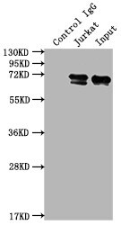
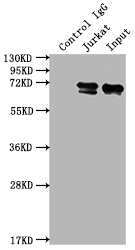
- Immunoprecipitating FUBP1 in Jurkat whole cell lysate. Lane 1: Rabbit control IgG Jurkat whole cell lysate. For western blotting, a HRP-conjugated Protein G antibody was used as the secondary antibody (1:2,000). Lane 2: FUBP1 Monoclonal Antibody (Product # MA5-38293) (2 µg) + Jurkat whole cell lysate (500 µg). Lane 3: Jurkat whole cell lysate (10 µg).
Supportive validation
- Submitted by
- Invitrogen Antibodies (provider)
- Main image

- Experimental details
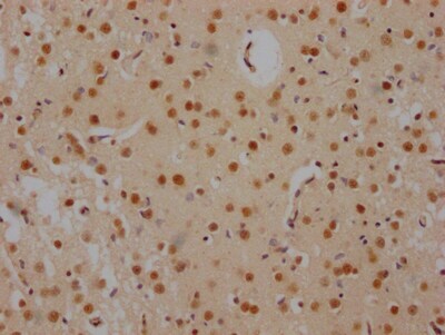
- Immunohistochemistry (Paraffin) analysis of FUBP1 in paraffin-embedded human brain tissue using FUBP1 Monoclonal Antibody (Product # MA5-38293) diluted at 1:100. After dewaxing and hydration, antigen retrieval was mediated by high pressure in a citrate buffer (pH 6.0). Section was blocked with 10% normal goat serum 30min at RT. Then primary antibody (1% BSA) was incubated at 4°C overnight. The primary is detected by a Goat anti-rabbit IgG polymer labeled by HRP and visualized using 0.05% DAB.
- Submitted by
- Invitrogen Antibodies (provider)
- Main image
- Experimental details
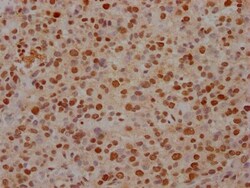
- Immunohistochemistry (Paraffin) analysis of FUBP1 in paraffin-embedded human glioma cancer using FUBP1 Monoclonal Antibody (Product # MA5-38293) diluted at 1:100. After dewaxing and hydration, antigen retrieval was mediated by high pressure in a citrate buffer (pH 6.0). Section was blocked with 10% normal goat serum 30min at RT. Then primary antibody (1% BSA) was incubated at 4°C overnight. The primary is detected by a Goat anti-rabbit IgG polymer labeled by HRP and visualized using 0.05% DAB.
Supportive validation
- Submitted by
- Invitrogen Antibodies (provider)
- Main image
- Experimental details
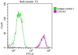
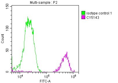
- Flow Cytometry analysis of FUBP1 in Jurkat cells using FUBP1 Monoclonal Antibody (Product # MA5-38293) at 1:50. The cells were fixed with 70% Ethylalcohol (18h) and then incubated in 10% normal goat serum to block non-specific protein-protein interactions followed by the antibody (1 µg/1x10^6 cells) for 1 h at 4°C. The secondary antibody used was FITC-conjugated goat anti-rabbit IgG (H+L) at 1:200 dilution for 30min at 4°C. Control antibody (green line) was Rabbit IgG (1 µg/1x10^6 cells) used under the same conditions. Acquisition of >10,000 events was performed.
- Submitted by
- Invitrogen Antibodies (provider)
- Main image

- Experimental details
- Flow Cytometry analysis of FUBP1 in Jurkat cells using FUBP1 Monoclonal Antibody (Product # MA5-38293) at 1:50. The cells were fixed with 70% Ethylalcohol (18h) and then incubated in 10% normal goat serum to block non-specific protein-protein interactions followed by the antibody (1 µg/1x10^6 cells) for 1 h at 4°C. The secondary antibody used was FITC-conjugated goat anti-rabbit IgG (H+L) at 1:200 dilution for 30min at 4°C. Control antibody (green line) was Rabbit IgG (1 µg/1x10^6 cells) used under the same conditions. Acquisition of >10,000 events was performed.

Supportive validation
- Submitted by
- Invitrogen Antibodies (provider)
- Main image

- Experimental details
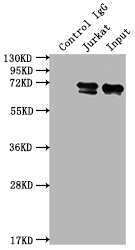
- Immunoprecipitating FUBP1 in Jurkat whole cell lysate. Lane 1: Rabbit control IgG Jurkat whole cell lysate. For western blotting, a HRP-conjugated Protein G antibody was used as the secondary antibody (1:2,000). Lane 2: FUBP1 Monoclonal Antibody (Product # MA5-38293) (2 µg) + Jurkat whole cell lysate (500 µg). Lane 3: Jurkat whole cell lysate (10 µg).