 Explore
 Validate
 Learn
Learn Western blot
 Immunocytochemistry
Antibody data
- Antibody Data
- Antigen structure
- References [0]
- Comments [0]
- Validations
- Immunocytochemistry [2]
- Immunoprecipitation [1]
- Immunohistochemistry [2]
- Chromatin Immunoprecipitation [1]
- Other assay [2]
Submit
Validation data
Reference
Comment
Report error
- Product number
- PA5-28061 - Provider product page

- Provider
- Invitrogen Antibodies
- Product name
- FUBP1 Polyclonal Antibody
- Antibody type
- Polyclonal
- Antigen
- Recombinant protein fragment
- Description
- Recommended positive controls: HeLa, HeLa nuclear extract, mouse brain, rat brain. Predicted reactivity: Mouse (99%), Rat (99%), Zebrafish (80%), Xenopus laevis (92%), Chicken (93%), Bovine (99%). Store product as a concentrated solution. Centrifuge briefly prior to opening the vial.
- Reactivity
- Human, Mouse, Rat
- Host
- Rabbit
- Isotype
- IgG
- Vial size
- 100 μL
- Concentration
- 0.92 mg/mL
- Storage
- Store at 4°C short term. For long term storage, store at -20°C, avoiding freeze/thaw cycles.
No comments: Submit comment
Supportive validation
- Submitted by
- Invitrogen Antibodies (provider)
- Main image
- Experimental details
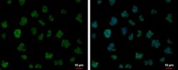
- FUBP1 Polyclonal Antibody detects FUBP1 protein at nucleus by immunofluorescent analysis. Sample: Jurkat cells were fixed in 4% paraformaldehyde at RT for 15 min. Green: FUBP1 protein stained by FUBP1 Polyclonal Antibody (Product # PA5-28061) diluted at 1:500. Blue: Hoechst 33342 staining.

- Submitted by
- Invitrogen Antibodies (provider)
- Main image

- Experimental details
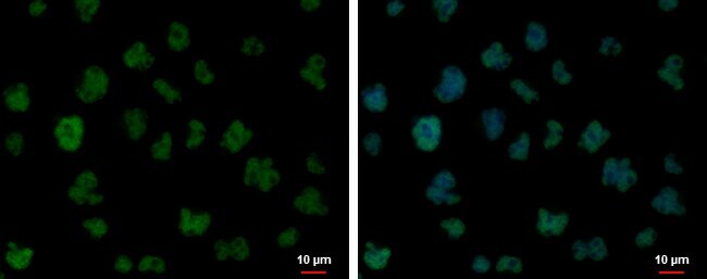
- FUBP1 Polyclonal Antibody detects FUBP1 protein at nucleus by immunofluorescent analysis. Sample: Jurkat cells were fixed in 4% paraformaldehyde at RT for 15 min. Green: FUBP1 protein stained by FUBP1 Polyclonal Antibody (Product # PA5-28061) diluted at 1:500. Blue: Hoechst 33342 staining.
Supportive validation
- Submitted by
- Invitrogen Antibodies (provider)
- Main image
- Experimental details
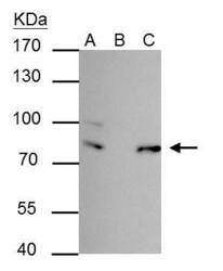
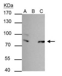
- FUBP-1 antibody immunoprecipitates FUBP-1 protein in IP experiments. IP Sample: 293T whole cell lysate/extract A : 30 µg whole cell lysate/extract of FUBP-1 protein expressing 293T cells B : Control with 2.5 µg of pre-immune rabbit IgG C : Immunoprecipitation of FUBP-1 by 2.5 µg of FUBP-1 antibody (Product # PA5-28061) 7.5% SDS-PAGE The immunoprecipitated FUBP-1 protein was detected by FUBP-1 antibody (Product # PA5-28061) diluted at 1:1,000. Anti-rabbit IgG (HRP) was used as a secondary reagent.
Supportive validation
- Submitted by
- Invitrogen Antibodies (provider)
- Main image
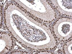
- Experimental details
- FUBP1 Polyclonal Antibody detects FUBP1 protein at nucleus on mouse testis by immunohistochemical analysis. Sample: Paraffin-embedded mouse testis. FUBP1 Polyclonal Antibody (Product # PA5-28061) dilution: 1:1,000. Antigen Retrieval: EDTA based buffer, pH 8.0, 15 min.
- Submitted by
- Invitrogen Antibodies (provider)
- Main image
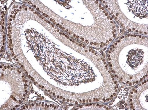
- Experimental details
- FUBP1 Polyclonal Antibody detects FUBP1 protein at nucleus on mouse testis by immunohistochemical analysis. Sample: Paraffin-embedded mouse testis. FUBP1 Polyclonal Antibody (Product # PA5-28061) dilution: 1:1,000. Antigen Retrieval: EDTA based buffer, pH 8.0, 15 min.
Supportive validation
- Submitted by
- Invitrogen Antibodies (provider)
- Main image
- Experimental details
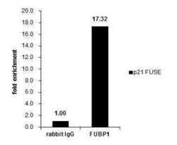
- Cross-linked ChIP was performed with HeLa chromatin extract and 5 µg of either control rabbit IgG or FUBP1 Polyclonal Antibody (Product # PA5-28061). The precipitated DNA was detected by PCR with primer set targeting to p21 FUSE.

Supportive validation
- Submitted by
- Invitrogen Antibodies (provider)
- Main image
- Experimental details
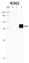
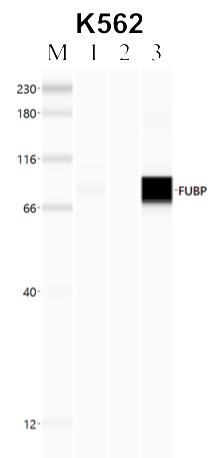
- RNA immunoprecipitation (RIP) western of FUBP1 was performed in K562 cells. Antigen-antibody complexes were formed by incubating approximately 500 µg whole cell lysate with 5 to 10 µL of polyclonal FUBP1 antibody (Product # PA5-28061) rotating 60 min at RT. The immune complexes were captured on 625 µg of anti-rabbit coated Dynabeads (Product # 11204D) and washed extensively. They were then eluted and analyzed using the Simple Western system using the same antibody as used in immunoprecipitation at a dilution of 1:25, followed by a 1:100 dilution of secondary antibody. Lane 1 is the input, lane 2 no antibody IP and lane 3 is the target specific IP. Data courtesy of the Yeo lab as part of the ENCODE project.
- Submitted by
- Invitrogen Antibodies (provider)
- Main image
- Experimental details
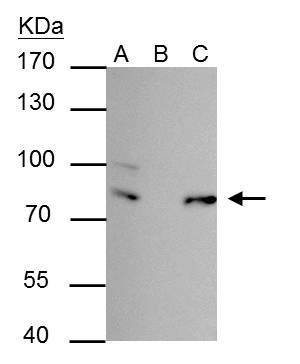
- FUBP-1 antibody immunoprecipitates FUBP-1 protein in IP experiments. IP Sample: 293T whole cell lysate/extract A : 30 µg whole cell lysate/extract of FUBP-1 protein expressing 293T cells B : Control with 2.5 µg of pre-immune rabbit IgG C : Immunoprecipitation of FUBP-1 by 2.5 µg of FUBP-1 antibody (Product # PA5-28061) 7.5% SDS-PAGE The immunoprecipitated FUBP-1 protein was detected by FUBP-1 antibody (Product # PA5-28061) diluted at 1:1,000. Anti-rabbit IgG (HRP) was used as a secondary reagent.
