Explore
 Validate
 Learn
Learn Western blot
Antibody data
- Antibody Data
- Antigen structure
- References [3]
- Comments [0]
- Validations
- Western blot [3]
- Immunocytochemistry [1]
- Immunoprecipitation [1]
- Immunohistochemistry [2]
- Chromatin Immunoprecipitation [1]
Submit
Validation data
Reference
Comment
Report error
- Product number
- GTX104579 - Provider product page

- Provider
- GeneTex
- Proper citation
- GeneTex Cat#GTX104579, RRID:AB_11165485
- Product name
- FUBP1 antibody
- Antibody type
- Polyclonal
- Reactivity
- Human, Mouse, Rat
- Host
- Rabbit
Submitted references Alternative splicing of U2AF1 reveals a shared repression mechanism for duplicated exons.
Exon-centric regulation of ATM expression is population-dependent and amenable to antisense modification by pseudoexon targeting.
FUBP1: a new protagonist in splicing regulation of the DMD gene.
Kralovicova J, Vorechovsky I
Nucleic acids research 2017 Jan 9;45(1):417-434
Nucleic acids research 2017 Jan 9;45(1):417-434
Exon-centric regulation of ATM expression is population-dependent and amenable to antisense modification by pseudoexon targeting.
Kralovicova J, Knut M, Cross NC, Vorechovsky I
Scientific reports 2016 Jan 6;6:18741
Scientific reports 2016 Jan 6;6:18741
FUBP1: a new protagonist in splicing regulation of the DMD gene.
Miro J, Laaref AM, Rofidal V, Lagrafeuille R, Hem S, Thorel D, Méchin D, Mamchaoui K, Mouly V, Claustres M, Tuffery-Giraud S
Nucleic acids research 2015 Feb 27;43(4):2378-89
Nucleic acids research 2015 Feb 27;43(4):2378-89
No comments: Submit comment
Supportive validation
- Submitted by
- GeneTex (provider)
- Main image
- Experimental details
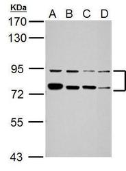
- Sample (30 ?g of whole cell lysate) A: Jurkat B: K562 C: THP-1 D: NCI-H929 7.5% SDS PAGE GTX104579 diluted at 1:5000 The HRP-conjugated anti-rabbit IgG antibody (GTX213110-01) was used to detect the primary antibody.
- Submitted by
- GeneTex (provider)
- Main image
- Experimental details
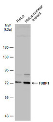
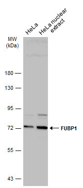
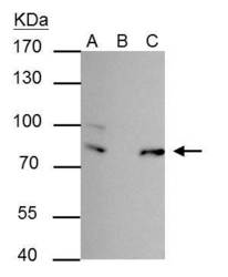
- Hela whole cell and nuclear extracts (30 ?g) were separated by 7.5% SDS-PAGE, and the membrane was blotted with FUBP1 antibody (GTX104579) diluted at 1:5000. The HRP-conjugated anti-rabbit IgG antibody (GTX213110-01) was used to detect the primary antibody.
- Submitted by
- GeneTex (provider)
- Main image
- Experimental details
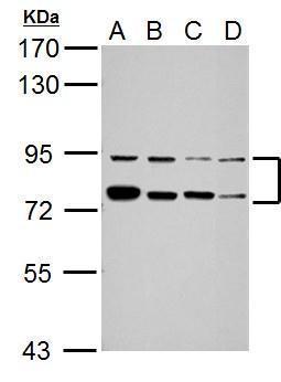
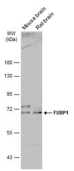
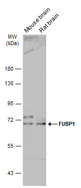
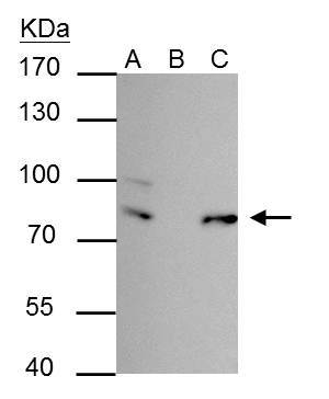
- Various tissue extracts (50 ?g) were separated by 7.5% SDS-PAGE, and the membrane was blotted with FUBP1 antibody (GTX104579) diluted at 1:5000. The HRP-conjugated anti-rabbit IgG antibody (GTX213110-01) was used to detect the primary antibody.
Supportive validation
- Submitted by
- GeneTex (provider)
- Main image
- Experimental details
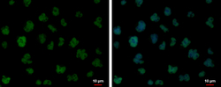
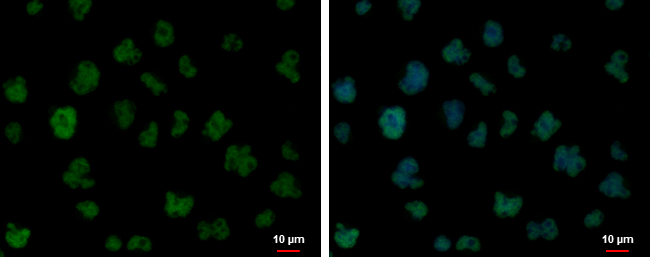
- FUBP1 antibody detects FUBP1 protein at nucleus by immunofluorescent analysis.Sample: Jurkat cells were fixed in 4% paraformaldehyde at RT for 15 min.Green: FUBP1 protein stained by FUBP1 antibody (GTX104579) diluted at 1:500.Blue: Hoechst 33342 staining.
Supportive validation
- Submitted by
- GeneTex (provider)
- Main image
- Experimental details
- FUBP-1 antibody immunoprecipitates FUBP-1 protein in IP experiments. IP Sample: 293T whole cell lysate/extract A : 30 £gg whole cell lysate/extract of FUBP-1 protein expressing 293T cells B : Control with 2.5 £gg of pre-immune rabbit IgG C : Immunoprecipitation of FUBP-1 by 2.5 £gg of FUBP-1 antibody (GTX104579) 7.5% SDS-PAGE The immunoprecipitated FUBP-1 protein was detected by FUBP-1 antibody (GTX1104579) diluted at 1 : 1000. EasyBlot anti-rabbit IgG (HRP) (GTX221666-01) was used as a secondary reagent.
Supportive validation
- Submitted by
- GeneTex (provider)
- Main image
- Experimental details
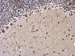
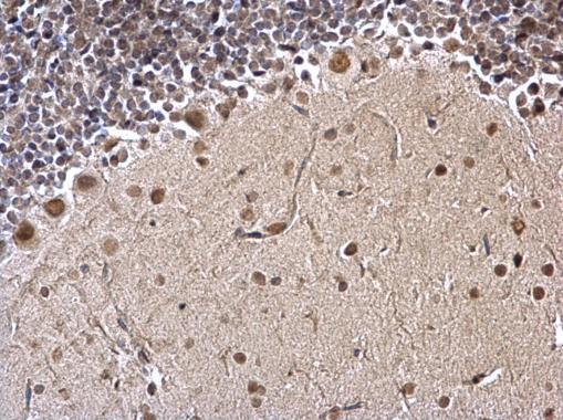
- FUBP1 antibody detects FUBP1 protein at nucleus on mouse hind brain by immunohistochemical analysis. Sample: Paraffin-embedded mouse hind brain. FUBP1 antibody (GTX104579) dilution: 1:1000.
- Submitted by
- GeneTex (provider)
- Main image
- Experimental details
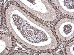
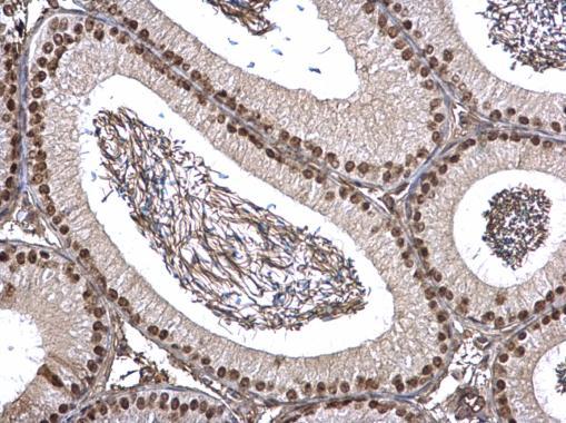
- FUBP1 antibody detects FUBP1 protein at nucleus on mouse testis by immunohistochemical analysis. Sample: Paraffin-embedded mouse testis. FUBP1 antibody (GTX104579) dilution: 1:1000.
Supportive validation
- Submitted by
- GeneTex (provider)
- Main image

- Experimental details
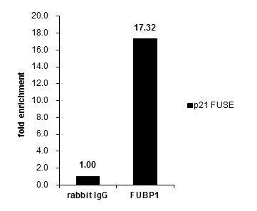
- Cross-linked ChIP was performed with HeLa chromatin extract and 5 £gg of either control rabbit IgG or anti-FUBP1 antibody. The precipitated DNA was detected by PCR with primer set targeting to p21 FUSE.