Explore
 Validate
 Learn
Learn Western blot
Antibody data
- Antibody Data
- Antigen structure
- References [0]
- Comments [0]
- Validations
- Western blot [1]
- Immunocytochemistry [1]
- Immunohistochemistry [1]
- Flow cytometry [1]
Submit
Validation data
Reference
Comment
Report error
- Product number
- 700241 - Provider product page

- Provider
- Invitrogen Antibodies
- Product name
- Phospho-AMPK beta-1 (Ser182) Recombinant Rabbit Monoclonal Antibody (9H26L42)
- Antibody type
- Monoclonal
- Antigen
- Synthetic peptide
- Reactivity
- Human
- Host
- Rabbit
- Isotype
- IgG
- Antibody clone number
- 9H26L42
- Vial size
- 100 µg
- Concentration
- 0.5 mg/mL
- Storage
- Store at 4°C short term. For long term storage, store at -20°C, avoiding freeze/thaw cycles.
No comments: Submit comment
Supportive validation
- Submitted by
- Invitrogen Antibodies (provider)
- Main image
- Experimental details
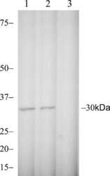
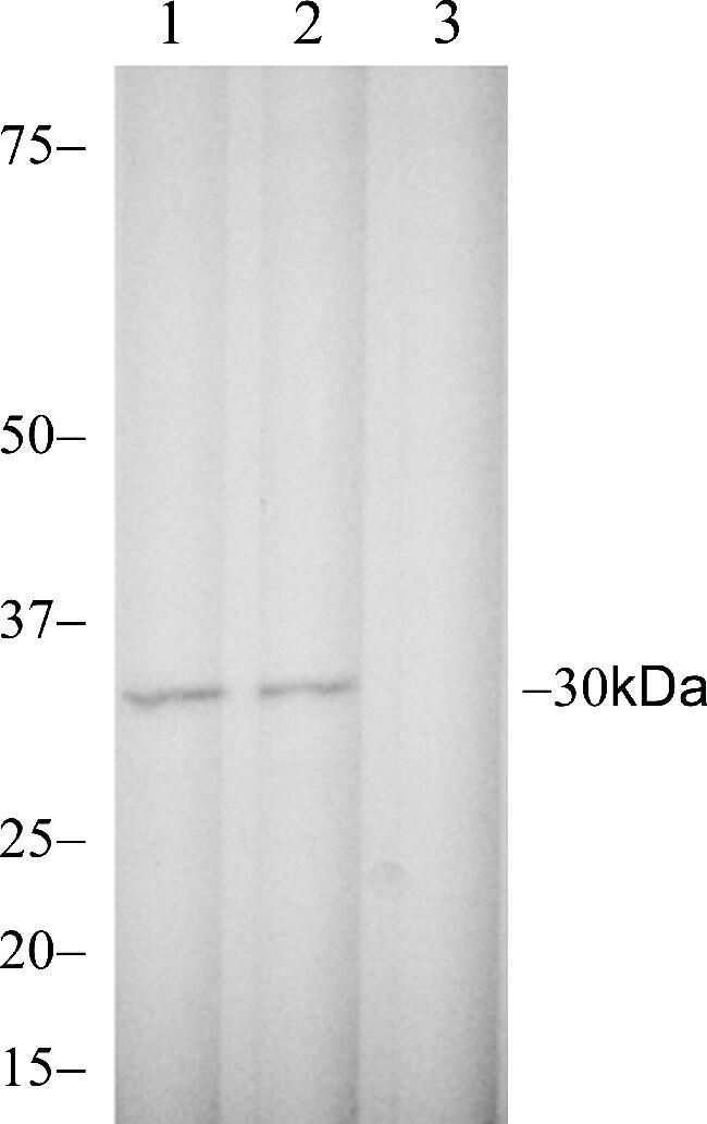
- Western blot analysis of Phospho-AMPK beta-1 pSer182 in Jurkat cell lysate using a Phospho-AMPK beta-1 pSer182 recombinant rabbit monoclonal antibody (Product # 700241) at a dilution of 2.5 µg/mL.
Supportive validation
- Submitted by
- Invitrogen Antibodies (provider)
- Main image

- Experimental details
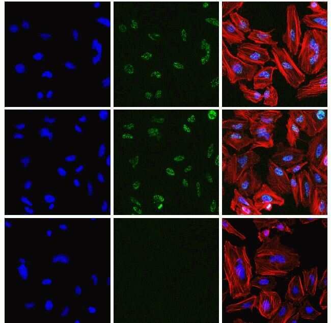
- Immunofluorescent analysis of Phospho-AMPK beta-1 pSer182 in HeLa cells using a Phospho-AMPK beta-1 pSer182 recombinant rabbit monoclonal antibody (Product # 700241) at a dilution of 2.5 µg/mL in the absence of peptide (top) and presence of immunogenic peptide (bottom), followed by detection using an Alexa Fluor 488-conjugated goat anti-rabbit secondary antibody at a dilution of 1:1000. Actin was stained with Alexa Fluor 568 Phalloidin (Product # A12380). Hoechst only (blue, left), AF488 signal only (green, middle) and composite image with Phalloidin (right).
Supportive validation
- Submitted by
- Invitrogen Antibodies (provider)
- Main image
- Experimental details
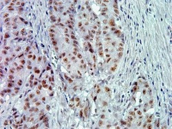
- Immunohistochemistry analysis of Phospho-AMPK beta-1 pSer182 in formalin-fixed, paraffin-embedded human colon carcimona using a Phospho-AMPK beta-1 pSer182 monoclonal antibody (Product # 700241) at a dilution of 5 µg/mL. Tissues were pretreated with EDTA and staining was visualized using DAB. Images were taken at a magnification of 40x. Results show nuclear staining in tumor cells.

Supportive validation
- Submitted by
- Invitrogen Antibodies (provider)
- Main image

- Experimental details
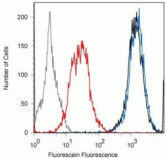
- Flow cytometry analysis of Phospho-AMPK beta-1 pSer182 in Jurkat cells using a Phospho-AMPK beta-1 pSer182 recombinant rabbit monoclonal antibody (Product # 700241) at a dilution of 0.5ug. Cells were fixed and permeabilized using FIX & PERM (Product # GAS004) reagent, and detection was performed using an Alexa Fluor 488 goat anti-rabbit IgG (black) compared to a control without primary antibody (gray). Pre-incubation with the phosphopeptide decreased the signal (red) whereas incubation with the non-phosphopeptide did not (blue).