Explore
 Validate
 Learn
Learn Western blot
 Immunocytochemistry
Antibody data
- Antibody Data
- Antigen structure
- References [0]
- Comments [0]
- Validations
- Immunocytochemistry [2]
- Immunohistochemistry [1]
- Flow cytometry [3]
Submit
Validation data
Reference
Comment
Report error
- Product number
- PA5-35341 - Provider product page

- Provider
- Invitrogen Antibodies
- Product name
- SCAP Polyclonal Antibody
- Antibody type
- Polyclonal
- Antigen
- Synthetic peptide
- Description
- This antibody is predicted to react with bovine, mouse, porcine and rat based on sequence homology.
- Reactivity
- Human, Mouse, Rat
- Host
- Rabbit
- Isotype
- IgG
- Vial size
- 400 μL
- Concentration
- 0.48 mg/mL
- Storage
- Store at 4°C short term. For long term storage, store at -20°C, avoiding freeze/thaw cycles.
No comments: Submit comment
Supportive validation
- Submitted by
- Invitrogen Antibodies (provider)
- Main image

- Experimental details
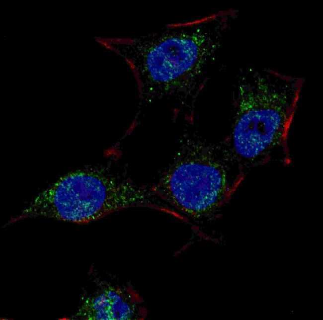
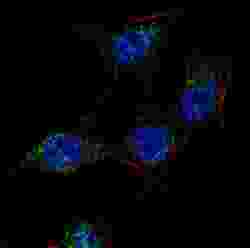
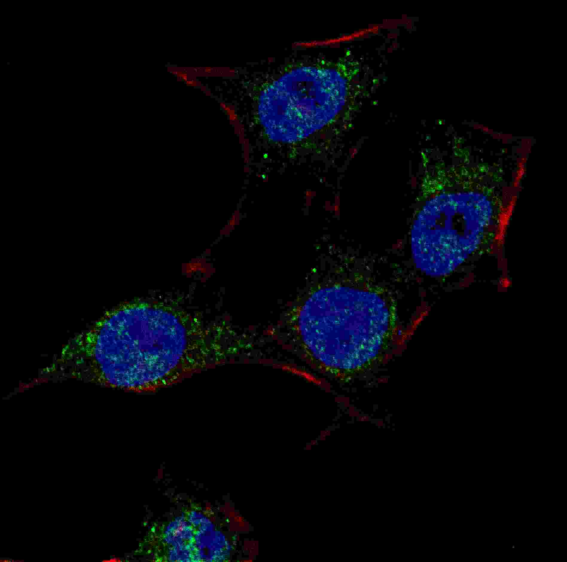
- Immunofluorescent analysis of SCAP in Hela cells. Hela cells were fixed with 4% PFA (20 min), permeabilized with Triton X-100 (0.2%, 30 min) and probed with a SCAP polyclonal antibody (Product # PA5-35341) at a dilution of 1:100 for 2 hours at room temperature, followed by detection using a fluorescent conjugated secondary antibody (green) (1:1000, 1h). Nuclei were stained with Hoechst 33342 (blue) (10 µg/mL, 5 min).
- Submitted by
- Invitrogen Antibodies (provider)
- Main image
- Experimental details
- Immunocytochemistry analysis of SCAP in HeLa cells. Samples were incubated with SCAP polyclonal antibody (Product # PA5-35341) using a dilution of 1:100 for 2 h at room temperature followed by Alexa Fluor® 488 conjugated donkey anti-rabbit at a dilution of 1:1,000 for 1hr. Cells were fixed with 4% PFA (20 min) and permeabilized with Triton X-100 (0.2%, 30 min). Nuclei were counterstained with Hoechst 33342 (blue) (10 µg/mL, 5 min).
Supportive validation
- Submitted by
- Invitrogen Antibodies (provider)
- Main image
- Experimental details
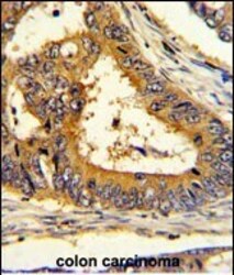
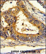
- Immunohistochemistry analysis of SCAP in formalin-fixed and paraffin-embedded human colon carcinoma. Samples were incubated with SCAP polyclonal antibody (Product # PA5-35341) which was peroxidase-conjugated to the secondary antibody, followed by DAB staining. This data demonstrates the use of this antibody for immunohistochemistry; clinical relevance has not been evaluated.
Supportive validation
- Submitted by
- Invitrogen Antibodies (provider)
- Main image

- Experimental details
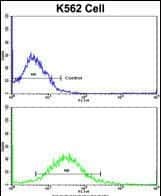
- Flow cytometry analysis of SCAP in K562 cells (bottom) compared to a negative control (top) using a SCAP polyclonal antibody (Product # PA5-35341) followed by detection using a FITC-conjugated goat-anti-rabbit secondary antibody.
- Submitted by
- Invitrogen Antibodies (provider)
- Main image
- Experimental details
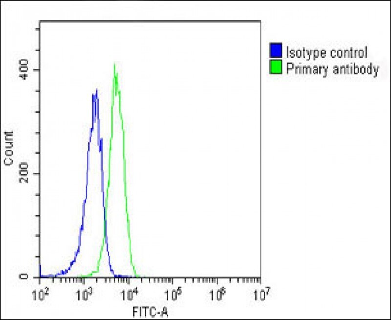
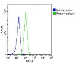
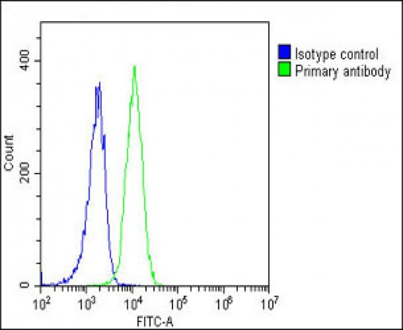
- Flow cytometry of (overlay histogram) of SCAP in K562 cells (green line). Samples were incubated with SCAP polyclonal antibody (Product # PA5-35341) using a dilution of 1:25 dilution for 60 min at 37°C followed by Goat-Anti-Rabbit IgG, DyLight® 488 Conjugated Highly Cross-Adsorbed at 1:200 dilution for 40 min at 37°C. The cells were fixed with 2% paraformaldehyde (10 min) and then permeabilized with 90% methanol for 10 min. The cells were then incubated in 2% bovine serum albumin to block non-specific protein-protein interactions followed by the primary antibody. Isotype control antibody (blue line) was rabbit IgG1 (1 μg/1x10^6 cells) used under the same conditions. Acquisition of >10, 000 events was performed.
- Submitted by
- Invitrogen Antibodies (provider)
- Main image
- Experimental details
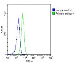
- Flow cytometry of (overlay histogram) of SCAP in K562 cells (green line). Samples were incubated with SCAP polyclonal antibody (Product # PA5-35341) using a dilution of 1:25 dilution for 60 min at 37°C followed by Goat-Anti-Rabbit IgG, DyLight® 488 Conjugated Highly Cross-Adsorbed at 1:200 dilution for 40 min at 37°C. The cells were fixed with 2% paraformaldehyde (10 min) and then permeabilized with 90% methanol for 10 min. The cells were then incubated in 2% bovine serum albumin to block non-specific protein-protein interactions followed by the primary antibody. Isotype control antibody (blue line) was rabbit IgG1 (1 μg/1x10^6 cells) used under the same conditions. Acquisition of >10, 000 events was performed.