 Explore
 Validate
 Learn
Learn Western blot
Antibody data
- Antibody Data
- Antigen structure
- References [2]
- Comments [0]
- Validations
- Western blot [1]
- Immunohistochemistry [1]
- Other assay [2]
Submit
Validation data
Reference
Comment
Report error
- Product number
- PA1-16800 - Provider product page

- Provider
- Invitrogen Antibodies
- Product name
- NPC1L1 Polyclonal Antibody
- Antibody type
- Polyclonal
- Antigen
- Synthetic peptide
- Description
- The target sequence has 93% sequence homology with human.
- Reactivity
- Human, Mouse, Rat, Porcine
- Host
- Rabbit
- Isotype
- IgG
- Vial size
- 100 μL
- Concentration
- 1 mg/mL
- Storage
- 4°C, do not freeze
Submitted references Novel Lactobacillus reuteri HI120 Affects Lipid Metabolism in C57BL/6 Obese Mice.
Caco-2 Cells for Measuring Intestinal Cholesterol Transport - Possibilities and Limitations.
Sun Y, Tang Y, Hou X, Wang H, Huang L, Wen J, Niu H, Zeng W, Bai Y
Frontiers in veterinary science 2020;7:560241
Frontiers in veterinary science 2020;7:560241
Caco-2 Cells for Measuring Intestinal Cholesterol Transport - Possibilities and Limitations.
Hiebl V, Schachner D, Ladurner A, Heiss EH, Stangl H, Dirsch VM
Biological procedures online 2020;22:7
Biological procedures online 2020;22:7
No comments: Submit comment
Supportive validation
- Submitted by
- Invitrogen Antibodies (provider)
- Main image
- Experimental details
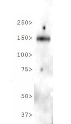
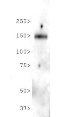
- Western blot analysis of NPC1L1 in lysate of human small intestine tissue. Samples were incubated in NPC1L1 polyclonal antibody (Product # PA1-16800) using a dilution of 2 µg/mL.
Supportive validation
- Submitted by
- Invitrogen Antibodies (provider)
- Main image
- Experimental details
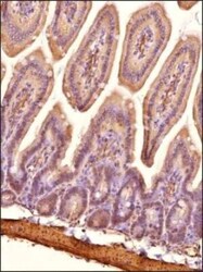
- Immunohistochemical analysis of NPC1L1 in formalin-fixed paraffin-embedded tissue section of mouse small intestine. Samples were incubated in NPC1L1 polyclonal antibody (Product # PA1-16800) using a dilution of 1:200 followed by HRP labeled anti-rabbit secondary antibody and DAB reagent. Nuclei of cells were counter-stained with hematoxylin.

Supportive validation
- Submitted by
- Invitrogen Antibodies (provider)
- Main image
- Experimental details
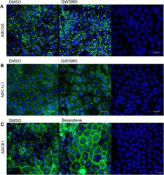
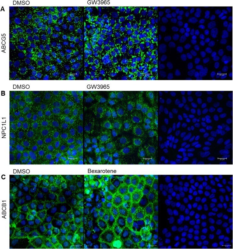
- Fig. 6 a-c Confocal laser scanning microscopy for the detection of ABCG5, NPC1L1 and ABCB1. Staining was carried out using Protocol #6. Cell nuclei are shown in blue (DAPI) and ABCG5, NPC1L1 or ABCB1 in green (fluorescently labelled antibody). Representative images of ABCG5 ( a ), treated with the vehicle control (DMSO 0.1%, left image) or GW3965 (LXR agonist, 10 muM, middle image). Representative images of NPC1L1 ( b ), treated with the vehicle control (DMSO 0.1%, left image) or GW3965 (LXR agonist, 10 muM, middle image). Representative images of ABCB1 ( c ), treated with the vehicle control (DMSO 0.1%, left image) or bexarotene (RXR agonist, 5 muM, middle image). The image on the right ( a-c ) is the respective control only stained with DAPI and the secondary antibody, showing the unspecific binding of the secondary antibody. Shown are maximum projections of the top views
- Submitted by
- Invitrogen Antibodies (provider)
- Main image
- Experimental details
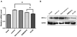
- Figure 8 mRNA and protein expression levels of NPC1L1 in all groups. (A) qRT-PCR results. (B) Western blotting results. * p < 0.05.
