Explore
 Validate
 Learn
LearnMA5-32618
antibody from Invitrogen Antibodies
Targeting: TNFRSF1B
CD120b, p75, TNF-R-II, TNF-R75, TNFBR, TNFR2, TNFR80
 Western blot
 Immunocytochemistry
Antibody data
- Antibody Data
- Antigen structure
- References [2]
- Comments [0]
- Validations
- Immunocytochemistry [7]
- Immunoprecipitation [1]
- Immunohistochemistry [5]
- Flow cytometry [3]
- Other assay [1]
Submit
Validation data
Reference
Comment
Report error
- Product number
- MA5-32618 - Provider product page

- Provider
- Invitrogen Antibodies
- Product name
- TNFR2 Recombinant Rabbit Monoclonal Antibody (JM113-01)
- Antibody type
- Monoclonal
- Antigen
- Synthetic peptide
- Description
- Recombinant rabbit monoclonal antibodies are produced using in vitro expression systems. The expression systems are developed by cloning in the specific antibody DNA sequences from immunoreactive rabbits. Then, individual clones are screened to select the best candidates for production. The advantages of using recombinant rabbit monoclonal antibodies include: better specificity and sensitivity, lot-to-lot consistency, animal origin-free formulations, and broader immunoreactivity to diverse targets due to larger rabbit immune repertoire.
- Reactivity
- Human, Mouse, Rat
- Host
- Rabbit
- Isotype
- IgG
- Antibody clone number
- JM113-01
- Vial size
- 100 μL
- Concentration
- 1 mg/mL
- Storage
- Store at 4°C short term. For long term storage, store at -20°C, avoiding freeze/thaw cycles.
Submitted references Tumor necrosis factor receptor-1 is selectively sequestered into Schwann cell extracellular vesicles where it functions as a TNFα decoy.
Upregulated tumor necrosis factor-α transcriptome and proteome in adipose tissue-derived mesenchymal stem cells from pigs with metabolic syndrome.
Sadri M, Hirosawa N, Le J, Romero H, Martellucci S, Kwon HJ, Pizzo D, Ohtori S, Gonias SL, Campana WM
Glia 2022 Feb;70(2):256-272
Glia 2022 Feb;70(2):256-272
Upregulated tumor necrosis factor-α transcriptome and proteome in adipose tissue-derived mesenchymal stem cells from pigs with metabolic syndrome.
Pawar AS, Eirin A, Tang H, Zhu XY, Lerman A, Lerman LO
Cytokine 2020 Mar 30;130:155080
Cytokine 2020 Mar 30;130:155080
No comments: Submit comment
Supportive validation
- Submitted by
- Invitrogen Antibodies (provider)
- Main image
- Experimental details
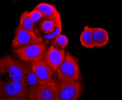
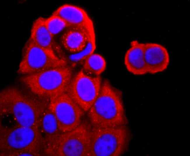
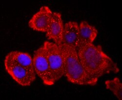
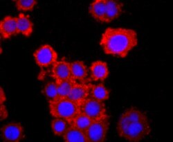
- Immunocytochemical analysis of TNFR2 in MCF-7 cells using a TNFR2 Monoclonal antibody (Product # MA5-32618) as seen in red. The nuclear counter stain is DAPI (blue). Cells were fixed in paraformaldehyde, permeabilised with 0.25% Triton X100/PBS.
- Submitted by
- Invitrogen Antibodies (provider)
- Main image
- Experimental details
- Immunocytochemical analysis of TNFR2 in Hela cells using a TNFR2 Monoclonal antibody (Product # MA5-32618) as seen in red. The nuclear counter stain is DAPI (blue). Cells were fixed in paraformaldehyde, permeabilised with 0.25% Triton X100/PBS.

- Submitted by
- Invitrogen Antibodies (provider)
- Main image
- Experimental details
- Immunocytochemical analysis of TNFR2 in SW480 cells using a TNFR2 Monoclonal antibody (Product # MA5-32618) as seen in red. The nuclear counter stain is DAPI (blue). Cells were fixed in paraformaldehyde, permeabilised with 0.25% Triton X100/PBS.

- Submitted by
- Invitrogen Antibodies (provider)
- Main image

- Experimental details
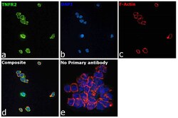
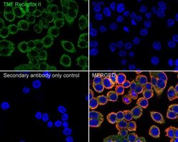
- Immunofluorescence analysis of Tumor necrosis factor receptor superfamily member 1B was performed using 70% confluent log phase THP-1 cells. The cells were fixed with 4% paraformaldehyde for 10 minutes, permeabilized with 0.1% Triton™ X-100 for 15 minutes, and blocked with 2% BSA for 45 minutes at room temperature. The cells were labeled with TNFR2 Recombinant Rabbit Monoclonal Antibody (JM113-01) (Product # MA5-32618) at 1:100 in 0.1% BSA, incubated at 4 degree celsius overnight and then labeled with Donkey anti-Rabbit IgG (H+L) Highly Cross-Adsorbed Secondary Antibody, Alexa Fluor Plus 488 (Product # A32790), (1:2000), for 45 minutes at room temperature (Panel a: Green). Nuclei (Panel b:Blue) were stained with ProLong™ Diamond Antifade Mountant with DAPI (Product # P36962). F-actin (Panel c: Red) was stained with Rhodamine Phalloidin (Product # R415, 1:300). Panel d represents the merged image showing Plasma Membrane localization. Panel e represents control cells with no primary antibody to assess background. The images were captured at 60X magnification.

- Submitted by
- Invitrogen Antibodies (provider)
- Main image
- Experimental details
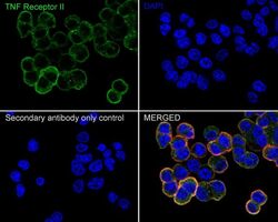
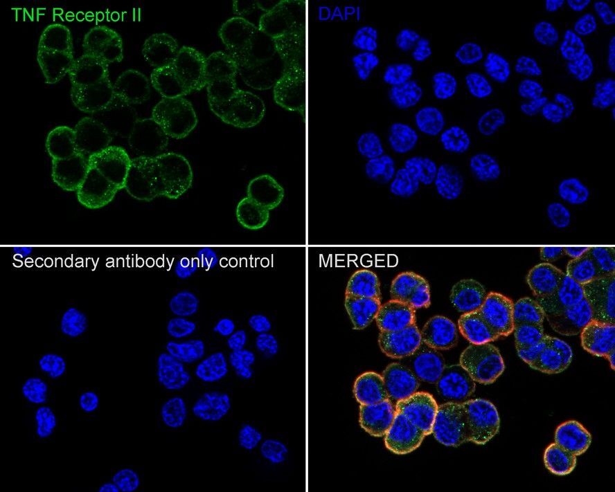
- Immunocytochemistry analysis of TNFR2 in PC-12 cells. Cells were fixed in 4% paraformaldehyde for 20 minutes at room temperature, permeabilized with 0.1% Triton X-100 in PBS for 5 minutes at room temperature, then blocked with 1% BSA in 10% negative goat serum for 1 hour at room temperature. Cells were then incubated with TNFR2 monoclonal antibody (Product # MA5-32618) using a dilution of 1:100 in 1% BSA in PBST overnight at 4 °C. Goat Anti-Rabbit IgG H&L (iFluor™ 488) was used as the secondary antibody at a 1:1,000 dilution. PBS instead of the primary antibody was used as the secondary antibody only control. Nuclear DNA was labelled in blue with DAPI. Beta tubulin (red) was stained at 1:100 dilution overnight at 4°C. Goat Anti-Mouse IgG H&L (iFluor™ 594) was used as the secondary antibody at a 1:1,000 dilution.
- Submitted by
- Invitrogen Antibodies (provider)
- Main image
- Experimental details
- Immunocytochemistry analysis of TNFR2 in RAW264.7 cells. Cells were fixed in 4% paraformaldehyde for 20 minutes at room temperature, permeabilized with 0.1% Triton X-100 in PBS for 5 minutes at room temperature, then blocked with 1% BSA in 10% negative goat serum for 1 hour at room temperature. Cells were then incubated with TNFR2 monoclonal antibody (Product # MA5-32618) using a dilution of 1:100 in 1% BSA in PBST overnight at 4 °C. Followed by Goat Anti-Rabbit IgG H&L (iFluor™ 488) at a 1:1,000 dilution. PBS instead of the primary antibody was used as the secondary antibody only control. Nuclear DNA was labelled in blue with DAPI. Beta tubulin (red) was stained at 1:100 dilution overnight at 4°C. Goat Anti-Mouse IgG H&L (iFluor™ 594) was used as the secondary antibody at a 1:1,000 dilution.

Supportive validation
- Submitted by
- Invitrogen Antibodies (provider)
- Main image
- Experimental details
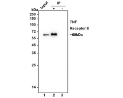
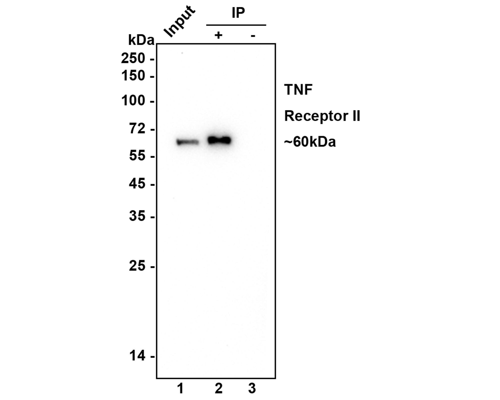
- Immunoprecipitation of TNFR2 in 0.2 mg Jurkat cell lysate. Samples were incubated with TNFR2 monoclonal antibody (Product # MA5-32618) at 2 µg/25 µL agarose. Western blot was performed from the immunoprecipitate using TNFR2 monoclonal antibody at a 1:1,000 dilution. Anti-Rabbit IgG for IP Nano-secondary antibody at a 1:5,000 dilution was used for 1 hour at room temperature. Lane 1: Jurkat cell lysate (input); Lane 2: TNFR2 antibody IP in Jurkat cell lysate; Lane 3: Rabbit IgG instead of primary in Jurkat cell lysate. Blocking/Dilution buffer: 5% NFDM/TBST. Exposure time: 3 minutes.
Supportive validation
- Submitted by
- Invitrogen Antibodies (provider)
- Main image
- Experimental details
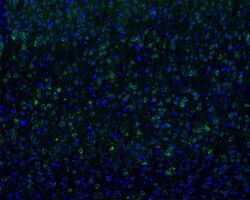
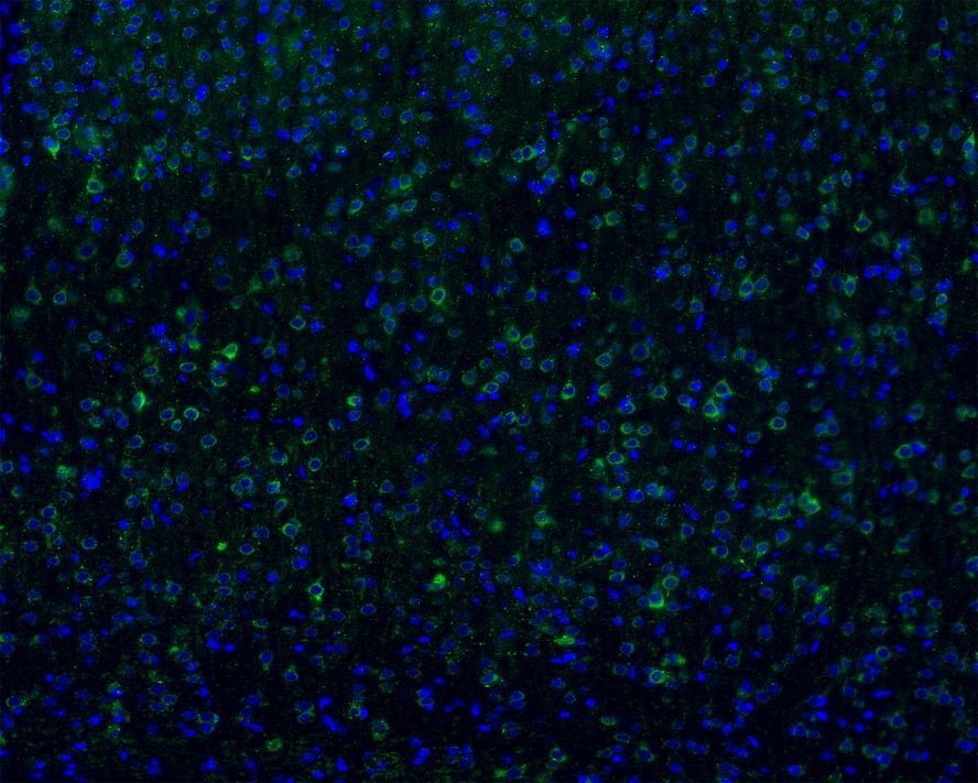
- Immunohistochemistry analysis of TNFR2 in frozen mouse cerebral cortex tissue. The tissues were blocked in 3% BSA for 30 minutes at room temperature, washed with PBS, and then probed with TNFR2 monoclonal antibody (Product # MA5-32618) using a dilution of 1:100 overnight at 4°C, washed with PBS. Followed by Goat Anti-Rabbit IgG H&L (Alexa Fluor® 488) at a 1:200 dilution. Nuclei were counterstained with DAPI (blue). Image acquisition was performed with KFBIO KF-FL-400 Scanner.
- Submitted by
- Invitrogen Antibodies (provider)
- Main image

- Experimental details
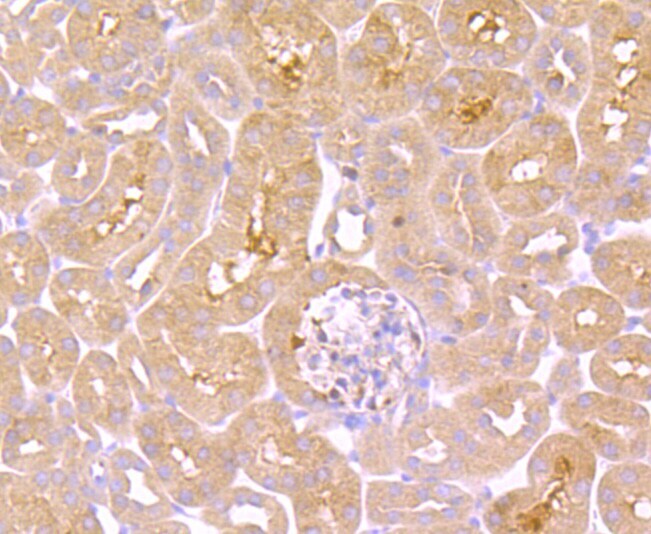
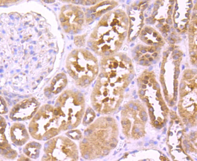
- Immunohistochemical analysis of TNFR2 of paraffin-embedded Mouse kidney tissue using a TNFR2 Monoclonal antibody (Product #MA5-32618). Counter stained with hematoxylin.
- Submitted by
- Invitrogen Antibodies (provider)
- Main image

- Experimental details
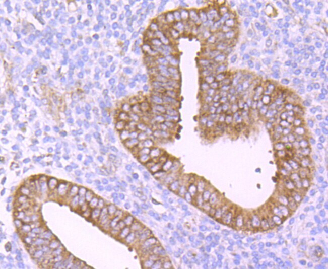
- Immunohistochemical analysis of TNFR2 of paraffin-embedded Human uterus tissue using a TNFR2 Monoclonal antibody (Product #MA5-32618). Counter stained with hematoxylin.
- Submitted by
- Invitrogen Antibodies (provider)
- Main image
- Experimental details
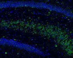
- Immunohistochemistry analysis of TNFR2 in frozen mouse hippocampus tissue. The tissues were blocked in 3% BSA for 30 minutes at room temperature, washed with PBS, and then probed with TNFR2 monoclonal antibody (Product # MA5-32618) using a dilution of 1:100 overnight at 4°C, washed with PBS. Followed by Goat Anti-Rabbit IgG H&L (Alexa Fluor® 488) at a 1:200 dilution. Nuclei were counterstained with DAPI (blue). Image acquisition was performed with KFBIO KF-FL-400 Scanner.

- Submitted by
- Invitrogen Antibodies (provider)
- Main image

- Experimental details
- Immunohistochemical analysis of TNFR2 of paraffin-embedded Human kidney tissue using a TNFR2 Monoclonal antibody (Product #MA5-32618). Counter stained with hematoxylin.
Supportive validation
- Submitted by
- Invitrogen Antibodies (provider)
- Main image

- Experimental details
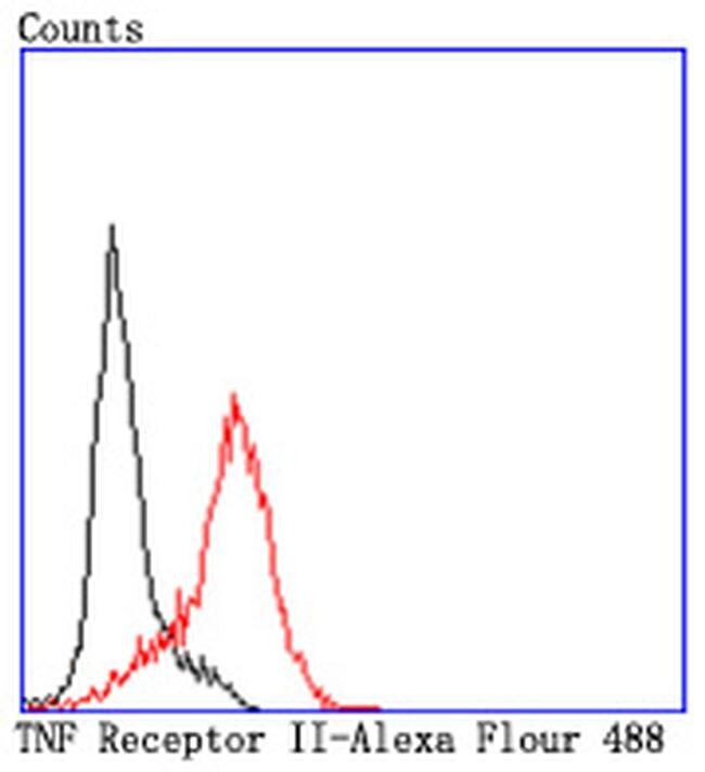
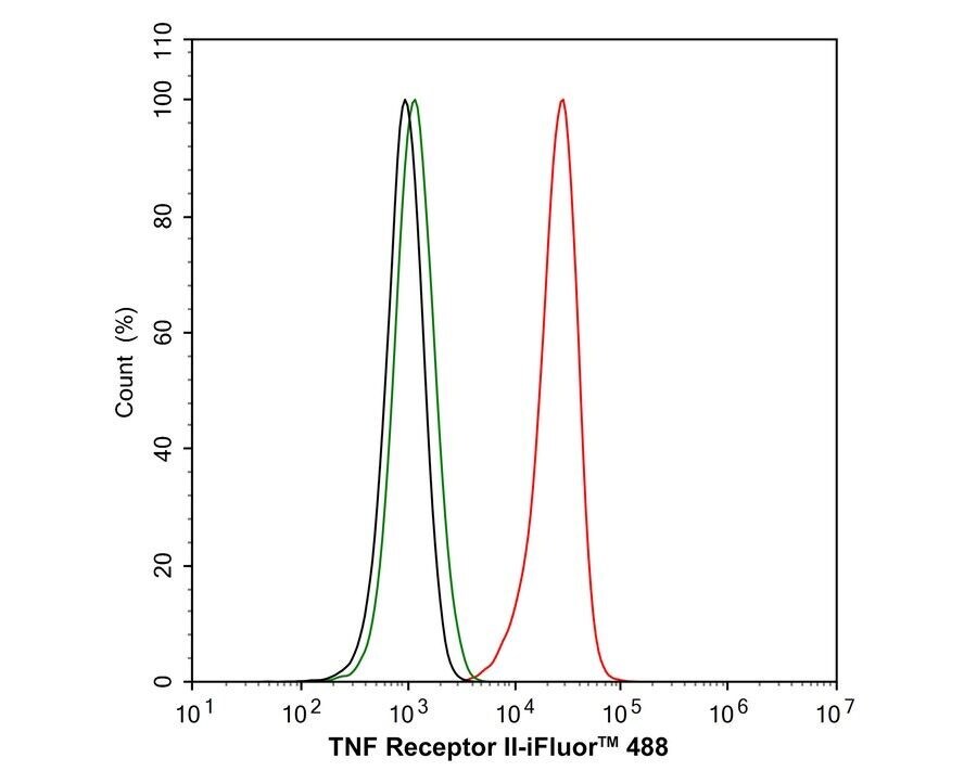
- Flow Cytometric analysis of TNFR2 in HL-60 cells using a TNFR2 Monoclonal Antibody (Product # MA5-32618) at a dilution of 1:50, as seen in red compared with an unlabelled control (cells without incubation with primary antibody; black). Alexa Fluor 488-conjugated goat anti rabbit IgG was used as the secondary antibody.
- Submitted by
- Invitrogen Antibodies (provider)
- Main image
- Experimental details
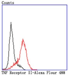
- Flow Cytometric analysis of TNFR2 in HL-60 cells using a TNFR2 Monoclonal Antibody (Product # MA5-32618) at a dilution of 1:50, as seen in red compared with an unlabelled control (cells without incubation with primary antibody; black). Alexa Fluor 488-conjugated goat anti rabbit IgG was used as the secondary antibody.

- Submitted by
- Invitrogen Antibodies (provider)
- Main image
- Experimental details
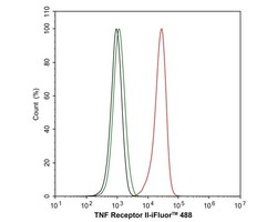
- Flow cytometry analysis of TNFR2 in RAW264.7 cells. Cells were fixed and permeabilized, then stained with TNFR2 monoclonal antibody (Product # MA5-32618) using a dilution of 1 µg/mL (red) compared with Rabbit IgG Isotype Control (green). After incubation of the primary antibody at 4°C for an hour, the cells were stained with a iFluor™ 488 conjugate-Goat anti-Rabbit IgG Secondary antibody at a 1:1,000 dilution for 30 minutes at 4°C. Unlabeled sample was used as a control (cells without incubation with primary antibody; black).
Supportive validation
- Submitted by
- Invitrogen Antibodies (provider)
- Main image
- Experimental details
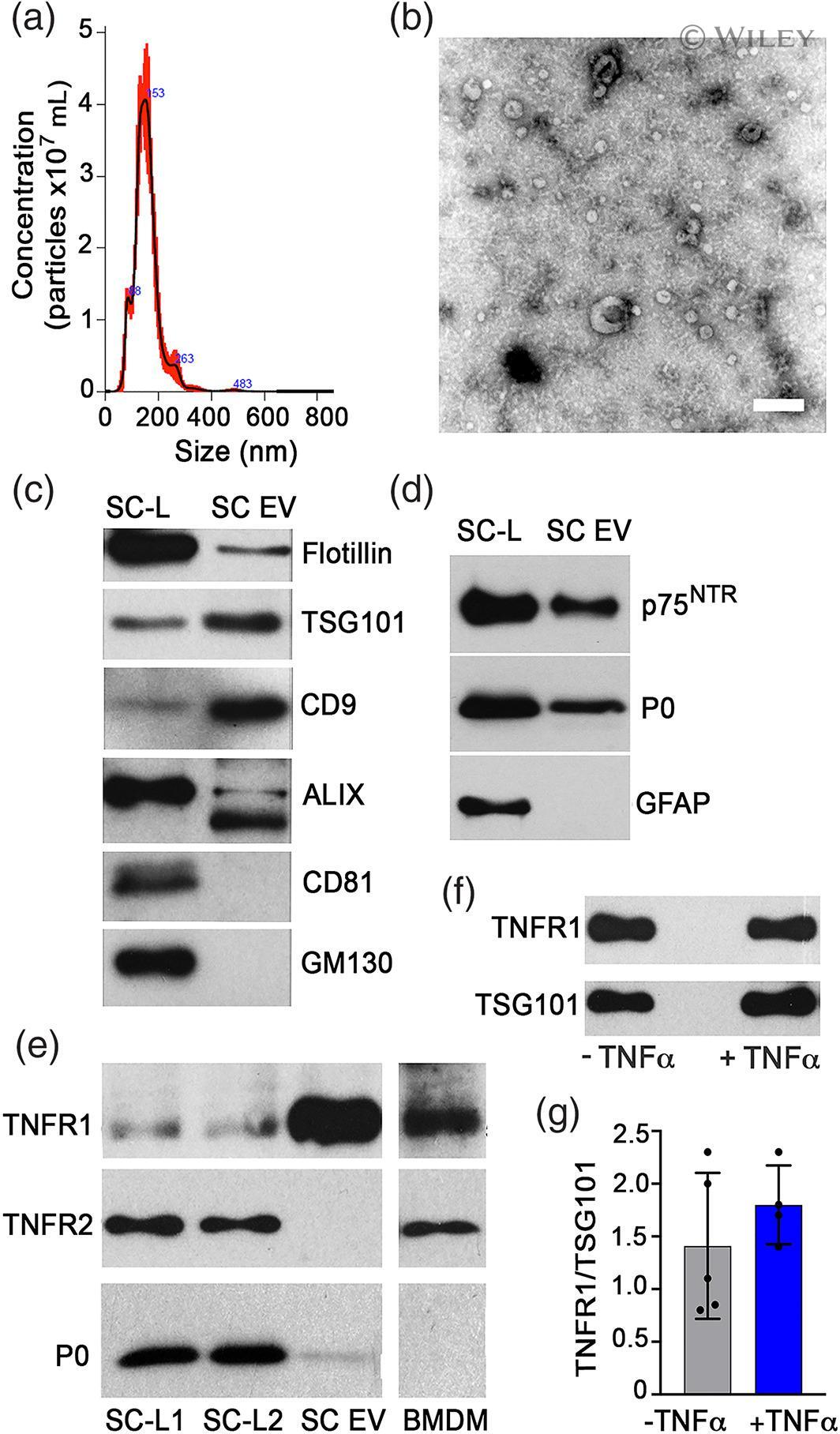
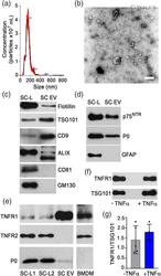
- 1 FIGURE TNFR1 is selectively sequestered into SC EVs. (a) SC EVs isolated by UC from conditioned media (CM) of primary cultured SCs were analyzed by NTA and had an average particle size of 153 +- 2 nm; n = 3 independent experiments. (b) Transmission electron microscopy (TEM) using negative staining of EVs. Note large crescent shaped particles and smaller particles. Scale bar, 200 nm. (c) Immunoblot analysis of whole SC lysates (SC-L) and SC-derived EVs to detect the exosome biomarkers, Flotillin-1, TSG101, CD9, ALIX, and CD81. GM130 is a Golgi biomarker not found in exosomes. (d) Immunoblot analysis to detect the SC biomarkers, p75 NTR and myelin protein zero, P0, and non-myelinating marker, GFAP. Images represent n = 3-5 independent experiments. (e) TNFalpha receptors, TNFR1 (55-kDa) and TNFR2 (65-kDa), and P0 in extracts of SCs cultured in complete medium (SC-L1) or in DMEM with 10% FBS depleted of EVs (SC-L2), in SC-EVs, and in bone marrow derived macrophages (BMDMs) (2 mug/lane) were determined by immunoblot analysis. (f) Immunoblot of TNFR1 levels in EVs derived from SCs treated with and without TNFalpha. TSG101 (44-kDa) shows load control in cells and presence in EVs. (g) Quantification of TNFR1 levels in SC EV immunoblots (2 mug). Data are expressed as mean +- SEM ; ( n = 4-5/group)