 Explore
 Validate
 Learn
Learn Western blot
 Flow cytometry
Antibody data
- Antibody Data
- Antigen structure
- References [0]
- Comments [0]
- Validations
- Western blot [3]
- Immunocytochemistry [1]
Submit
Validation data
Reference
Comment
Report error
- Product number
- GTX629694 - Provider product page

- Provider
- GeneTex
- Product name
- E-Cadherin antibody [GT358]
- Antibody type
- Monoclonal
- Reactivity
- Human
- Host
- Mouse
No comments: Submit comment
Supportive validation
- Submitted by
- GeneTex (provider)
- Main image

- Experimental details
- E-cadherin antibody [GT358] detects E-cadherin protein by Western blot analysis.A. 30 £gg MCF-7 whole cell lysate/extractB. 30 £gg MDA-MB-231 whole cell lysate/extract7.5 % SDS-PAGEE-cadherin antibody [GT358] (GTX629694) dilution: 1:1000

- Submitted by
- GeneTex (provider)
- Main image

- Experimental details
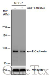
- Non-transfected (¡V) and transfected (+) MCF-7 whole cell extracts (30 ?g) were separated by 5% SDS-PAGE, and the membrane was blotted with E-Cadherin antibody [GT358] (GTX629694) diluted at 1:500. The HRP-conjugated anti-mouset IgG antibody (GTX213111-01) was used to detect the primary antibody.

- Submitted by
- GeneTex (provider)
- Main image
- Experimental details
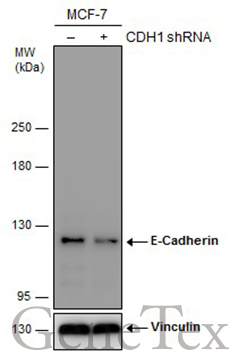
- Non-transfected (¡V) and transfected (+) MCF-7 whole cell extracts (30 ?g) were separated by 5% SDS-PAGE, and the membrane was blotted with E-Cadherin antibody [GT358] (GTX629694) diluted at 1:500. The HRP-conjugated anti-mouset IgG antibody (GTX213111-01) was used to detect the primary antibody.
Supportive validation
- Submitted by
- GeneTex (provider)
- Main image
- Experimental details
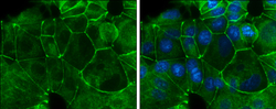
- E-Cadherin antibody [GT358] detects E-Cadherin protein at cell membrane by immunofluorescent analysis.Sample: MCF7 cells were fixed in ice-cold MeOH for 5 min.Green: E-Cadherin protein stained by E-Cadherin antibody [GT358] (GTX629694) diluted at 1:500.Blue: Hoechst 33342 staining.
