Explore
 Validate
 Learn
Learn Western blot
Antibody data
- Antibody Data
- Antigen structure
- References [10]
- Comments [0]
- Validations
- Western blot [2]
- Immunohistochemistry [1]
Submit
Validation data
Reference
Comment
Report error
- Product number
- MAB1838-100 - Provider product page

- Provider
- R&D Systems
- Product name
- Human E-Cadherin Antibody
- Antibody type
- Monoclonal
- Description
- Protein A or G purified from hybridoma culture supernatant. Detects human E-Cadherin in direct ELISAs and Western blots. In direct ELISAs and Western blots, no cross-reactivity with recombinant human (rh) Cadherin-8, rhCadherin-17, recombinant mouse E-Cadherin, rhN-Cadherin, rhP-Cadherin, or rhVE-Cadherin is observed.
- Reactivity
- Human
- Host
- Mouse
- Conjugate
- Unconjugated
- Antigen sequence
P12830- Isotype
- IgG
- Antibody clone number
- 180215
- Vial size
- 100 ug
- Concentration
- LYOPH
- Storage
- Use a manual defrost freezer and avoid repeated freeze-thaw cycles. 12 months from date of receipt, -20 to -70 °C as supplied. 1 month, 2 to 8 °C under sterile conditions after reconstitution. 6 months, -20 to -70 °C under sterile conditions after reconstitution.
Submitted references Neutrophil extracellular traps (NETs) contribute to pathological changes of ocular graft-vs.-host disease (oGVHD) dry eye: Implications for novel biomarkers and therapeutic strategies.
High content screening identifies monensin as an EMT-selective cytotoxic compound.
Modeling of Aniridia-Related Keratopathy by CRISPR/Cas9 Genome Editing of Human Limbal Epithelial Cells and Rescue by Recombinant PAX6 Protein.
Selective Laminin-Directed Differentiation of Human Induced Pluripotent Stem Cells into Distinct Ocular Lineages.
Zeylenone represses the progress of human prostate cancer by downregulating the Wnt/β‑catenin pathway.
Rose Bengal acetate photodynamic therapy (RBAc-PDT) induces exposure and release of Damage-Associated Molecular Patterns (DAMPs) in human HeLa cells.
Glycogene expression alterations associated with pancreatic cancer epithelial-mesenchymal transition in complementary model systems.
Intratumoral induction of CD103 triggers tumor-specific CTL function and CCR5-dependent T-cell retention.
Vascular endothelial growth factor-A stimulates Snail expression in breast tumor cells: implications for tumor progression.
Expression of mesenchyme-specific gene HMGA2 in squamous cell carcinomas of the oral cavity.
An S, Raju I, Surenkhuu B, Kwon JE, Gulati S, Karaman M, Pradeep A, Sinha S, Mun C, Jain S
The ocular surface 2019 Jul;17(3):589-614
The ocular surface 2019 Jul;17(3):589-614
High content screening identifies monensin as an EMT-selective cytotoxic compound.
Vanneste M, Huang Q, Li M, Moose D, Zhao L, Stamnes MA, Schultz M, Wu M, Henry MD
Scientific reports 2019 Feb 4;9(1):1200
Scientific reports 2019 Feb 4;9(1):1200
Modeling of Aniridia-Related Keratopathy by CRISPR/Cas9 Genome Editing of Human Limbal Epithelial Cells and Rescue by Recombinant PAX6 Protein.
Roux LN, Petit I, Domart R, Concordet JP, Qu J, Zhou H, Joliot A, Ferrigno O, Aberdam D
Stem cells (Dayton, Ohio) 2018 Sep;36(9):1421-1429
Stem cells (Dayton, Ohio) 2018 Sep;36(9):1421-1429
Selective Laminin-Directed Differentiation of Human Induced Pluripotent Stem Cells into Distinct Ocular Lineages.
Shibata S, Hayashi R, Okubo T, Kudo Y, Katayama T, Ishikawa Y, Toga J, Yagi E, Honma Y, Quantock AJ, Sekiguchi K, Nishida K
Cell reports 2018 Nov 6;25(6):1668-1679.e5
Cell reports 2018 Nov 6;25(6):1668-1679.e5
Zeylenone represses the progress of human prostate cancer by downregulating the Wnt/β‑catenin pathway.
Zeng S, Zhu B, Zeng J, Wu W, Jiang C
Molecular medicine reports 2018 Dec;18(6):5572-5578
Molecular medicine reports 2018 Dec;18(6):5572-5578
Rose Bengal acetate photodynamic therapy (RBAc-PDT) induces exposure and release of Damage-Associated Molecular Patterns (DAMPs) in human HeLa cells.
Panzarini E, Inguscio V, Fimia GM, Dini L
PloS one 2014;9(8):e105778
PloS one 2014;9(8):e105778
Glycogene expression alterations associated with pancreatic cancer epithelial-mesenchymal transition in complementary model systems.
Maupin KA, Sinha A, Eugster E, Miller J, Ross J, Paulino V, Keshamouni VG, Tran N, Berens M, Webb C, Haab BB
PloS one 2010 Sep 27;5(9):e13002
PloS one 2010 Sep 27;5(9):e13002
Intratumoral induction of CD103 triggers tumor-specific CTL function and CCR5-dependent T-cell retention.
Franciszkiewicz K, Le Floc'h A, Jalil A, Vigant F, Robert T, Vergnon I, Mackiewicz A, Benihoud K, Validire P, Chouaib S, Combadière C, Mami-Chouaib F
Cancer research 2009 Aug 1;69(15):6249-55
Cancer research 2009 Aug 1;69(15):6249-55
Vascular endothelial growth factor-A stimulates Snail expression in breast tumor cells: implications for tumor progression.
Wanami LS, Chen HY, Peiró S, García de Herreros A, Bachelder RE
Experimental cell research 2008 Aug 1;314(13):2448-53
Experimental cell research 2008 Aug 1;314(13):2448-53
Expression of mesenchyme-specific gene HMGA2 in squamous cell carcinomas of the oral cavity.
Miyazawa J, Mitoro A, Kawashiri S, Chada KK, Imai K
Cancer research 2004 Mar 15;64(6):2024-9
Cancer research 2004 Mar 15;64(6):2024-9
No comments: Submit comment
Supportive validation
- Submitted by
- R&D Systems (provider)
- Main image
- Experimental details
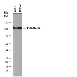
- Detection of Human E-Cadherin by Western Blot. Western blot shows lysates of A549 human lung carcinoma cell line and HepG2 human hepatocellular carcinoma cell line. PVDF membrane was probed with 0.5 µg/mL of Mouse Anti-Human E-Cadherin Monoclonal Antibody (Catalog # MAB1838) followed by HRP-conjugated Anti-Mouse IgG Secondary Antibody (Catalog # HAF018). A specific band was detected for E-Cadherin at approximately 110 kDa (as indicated). This experiment was conducted under reducing conditions and using Immunoblot Buffer Group 1.

- Submitted by
- R&D Systems (provider)
- Main image

- Experimental details
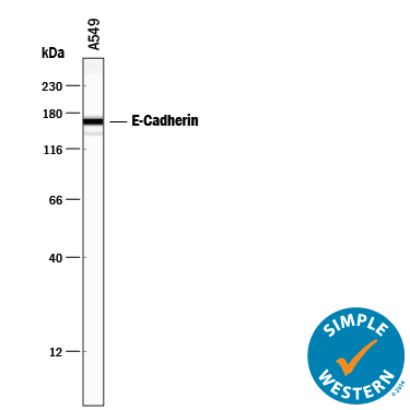
- Detection of Human E-Cadherin by Simple WesternTM. Simple Western lane view shows lysates of A549 human lung carcinoma cell line, loaded at 0.2 mg/mL. A specific band was detected for E-Cadherin at approximately 166 kDa (as indicated) using 5 µg/mL of Mouse Anti-Human E-Cadherin Monoclonal Antibody (Catalog # MAB1838) . This experiment was conducted under reducing conditions and using the 12-230 kDa separation system.
Supportive validation
- Submitted by
- R&D Systems (provider)
- Main image
- Experimental details
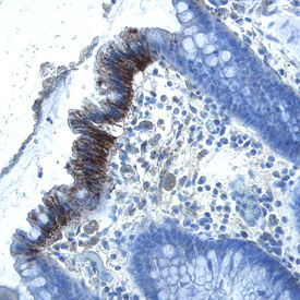
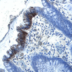
- E-Cadherin in Human Colon. E-Cadherin was detected in immersion fixed paraffin-embedded sections of human colon using Mouse Anti-Human E-Cadherin Monoclonal Antibody (Catalog # MAB1838) at 2 µg/mL overnight at 4 °C. Tissue was stained using the Anti-Mouse HRP-DAB Cell & Tissue Staining Kit (brown; Catalog # CTS002) and counterstained with hematoxylin (blue). Specific labeling was localized to the plasma membrane of epithelial cells. View our protocol for Chromogenic IHC Staining of Paraffin-embedded Tissue Sections.