 Explore
 Validate
 Learn
Learn Western blot
Antibody data
- Antibody Data
- Antigen structure
- References [0]
- Comments [0]
- Validations
- Western blot [2]
- Immunocytochemistry [2]
- Immunohistochemistry [1]
- Flow cytometry [1]
- Other assay [2]
Submit
Validation data
Reference
Comment
Report error
- Product number
- V3232 - Provider product page

- Provider
- NSJ Bioreagents
- Product name
- E-Cadherin Antibody
- Antibody type
- Monoclonal
- Description
- This highly specific E-Cadherin antibody is suitable for use in Flow cytometry/Immunofluorescence/Western blot/Immunohistochemistry applications with human, mouse and rat samples.
- Reactivity
- Human, Mouse, Rat
- Host
- Mouse
- Conjugate
- Unconjugated
- Antibody clone number
- 4A2
- Vial size
- 20 ug (with BSA and sodium azide), 100 ug (with BSA and sodium azide), 100 ug (without BSA or sodium azide), 7 ml IHC only format (if applicable)
- Concentration
- 0.2 mg/ml, 1 mg/ml
- Storage
- Store the E-Cadherin antibody at 2-8oC (with azide) or aliquot and store at -20oC or colder (without azide).
No comments: Submit comment
Supportive validation
- Submitted by
- NSJ Bioreagents (provider)
- Main image
- Experimental details
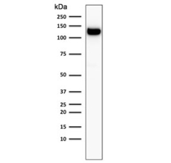
- Western blot testing of human MCF-7 cell lysate with E-Cadherin antibody (clone 4A2). Expected molecular weight: 135 kDa (precursor), 80-120 kDa (mature, depending on glycosylation level).

- Submitted by
- NSJ Bioreagents (provider)
- Main image
- Experimental details
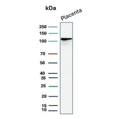
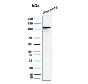
- Western blot testing of human placental tissue lysate with E-Cadherin antibody (clone 4A2). Expected molecular weight: 135 kDa (precursor), 80-120 kDa (mature, depending on glycosylation level).
Supportive validation
- Submitted by
- NSJ Bioreagents (provider)
- Main image
- Experimental details
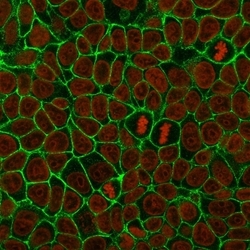
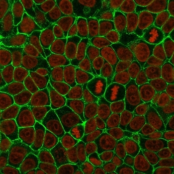
- Immunofluorescent staining of human MCF7 cells with E-Cadherin antibody (green, clone 4A2) and Reddot nuclear stain (red).
- Submitted by
- NSJ Bioreagents (provider)
- Main image

- Experimental details
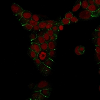
- Immunofluorescent staining of methanol-fixed human MCF7 cells with E-Cadherin antibody (green, clone 4A2) and Reddot nuclear stain (red).
Supportive validation
- Submitted by
- NSJ Bioreagents (provider)
- Main image

- Experimental details
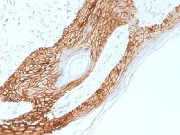
- IHC testing of FFPE human skin with E-Cadherin antibody (clone 4A2). Required HIER: boil tissue sections in pH 9 10mM Tris with 1mM EDTA for 10-20 min.
Supportive validation
- Submitted by
- NSJ Bioreagents (provider)
- Main image
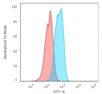
- Experimental details
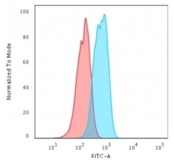
- Flow cytometry testing of human MCF7 cells with E-Cadherin antibody (clone 4A2); Red=isotype control, Blue= E-Cadherin antibody.
Supportive validation
- Submitted by
- NSJ Bioreagents (provider)
- Main image
- Experimental details
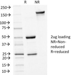
- SDS-PAGE Analysis of Purified, BSA-Free E-Cadherin Antibody (clone 4A2). Confirmation of Integrity and Purity of the Antibody.

- Submitted by
- NSJ Bioreagents (provider)
- Main image

- Experimental details
- SDS-PAGE analysis of purified, BSA-free E-Cadherin antibody (clone 4A2) as confirmation of integrity and purity.
