Explore
 Validate
 Learn
Learn Western blot
 Immunocytochemistry
Antibody data
- Antibody Data
- Antigen structure
- References [2]
- Comments [0]
- Validations
- Immunocytochemistry [6]
- Immunoprecipitation [2]
- Immunohistochemistry [8]
- Other assay [3]
Submit
Validation data
Reference
Comment
Report error
- Product number
- PA5-85088 - Provider product page

- Provider
- Invitrogen Antibodies
- Product name
- E-cadherin Polyclonal Antibody
- Antibody type
- Polyclonal
- Antigen
- Recombinant full-length protein
- Description
- Keep as concentrated solution. Predicted reactivity: Mouse (80%), Rat (83%), Dog (80%), Pig (83%), Bovine (82%). Positive Control: MCF-7, HT-29. Store product as a concentrated solution. Centrifuge briefly prior to opening the vial.
- Reactivity
- Human, Mouse, Rat, Zebrafish
- Host
- Rabbit
- Isotype
- IgG
- Vial size
- 100 μL
- Concentration
- 0.14 mg/mL
- Storage
- Store at 4°C short term. For long term storage, store at -20°C, avoiding freeze/thaw cycles.
Submitted references hsa_circ_0067514 suppresses gastric cancer progression and glycolysis via miR-654-3p/LATS2 axis.
Innate immune signaling in trophoblast and decidua organoids defines differential antiviral defenses at the maternal-fetal interface.
Jiang SF, Li RR
Neoplasma 2022 Sep;69(5):1079-1091
Neoplasma 2022 Sep;69(5):1079-1091
Innate immune signaling in trophoblast and decidua organoids defines differential antiviral defenses at the maternal-fetal interface.
Yang L, Semmes EC, Ovies C, Megli C, Permar S, Gilner JB, Coyne CB
eLife 2022 Aug 17;11
eLife 2022 Aug 17;11
No comments: Submit comment
Supportive validation
- Submitted by
- Invitrogen Antibodies (provider)
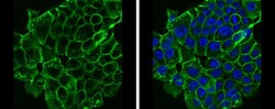
- Main image

- Experimental details
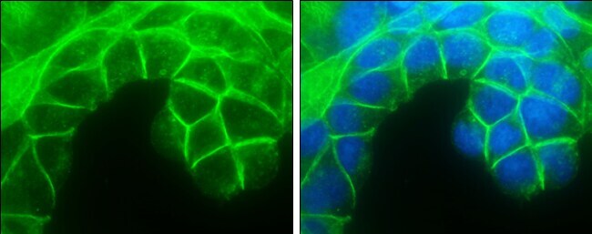
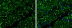
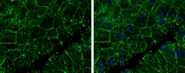
- Immunocytochemistry analysis of E-cadherin in 4% paraformaldehyde-fixed MCF7 cells using E-cadherin polyclonal antibody (Product # PA5-85088) at a dilution of 1:500. Sample was then incubated with Hoechst secondary antibody.
- Submitted by
- Invitrogen Antibodies (provider)
- Main image

- Experimental details
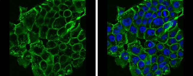
- Immunocytochemistry analysis of E-cadherin in 4% paraformaldehyde-fixed HCT 116 cells using E-cadherin polyclonal antibody (Product # PA5-85088) at a dilution of 1:500. Sample was then incubated with Hoechst secondary antibody.

- Submitted by
- Invitrogen Antibodies (provider)
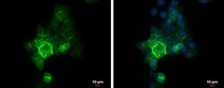
- Main image

- Experimental details
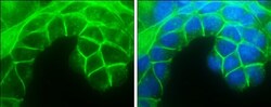
- Immunocytochemistry analysis of E-cadherin in 4% paraformaldehyde-fixed A431 cells using E-cadherin polyclonal antibody (Product # PA5-85088) at a dilution of 1:500. Sample was then incubated with Hoechst secondary antibody.
- Submitted by
- Invitrogen Antibodies (provider)
- Main image
- Experimental details
- Immunocytochemistry analysis of E-cadherin in 4% paraformaldehyde-fixed MCF7 cells using E-cadherin polyclonal antibody (Product # PA5-85088) at a dilution of 1:500. Sample was then incubated with Hoechst secondary antibody.

- Submitted by
- Invitrogen Antibodies (provider)
- Main image
- Experimental details
- Immunocytochemistry analysis of E-cadherin in 4% paraformaldehyde-fixed A431 cells using E-cadherin polyclonal antibody (Product # PA5-85088) at a dilution of 1:500. Sample was then incubated with Hoechst secondary antibody.

- Submitted by
- Invitrogen Antibodies (provider)
- Main image
- Experimental details
- Immunocytochemistry analysis of E-cadherin in 4% paraformaldehyde-fixed HCT 116 cells using E-cadherin polyclonal antibody (Product # PA5-85088) at a dilution of 1:500. Sample was then incubated with Hoechst secondary antibody.

Supportive validation
- Submitted by
- Invitrogen Antibodies (provider)
- Main image
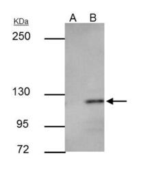
- Experimental details
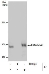
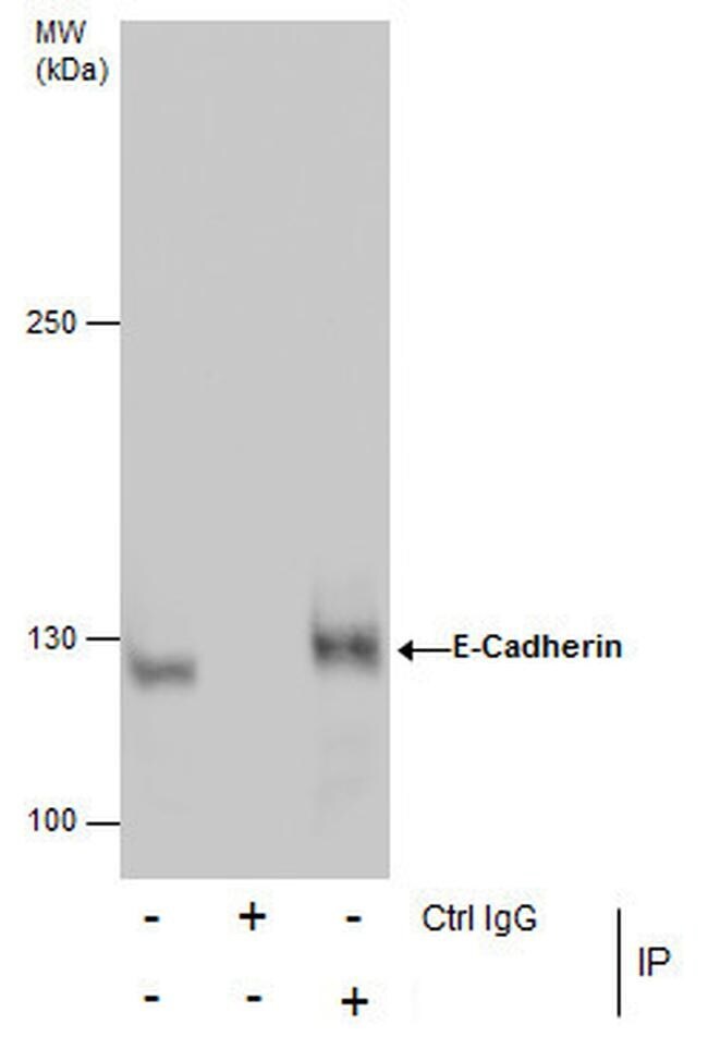
- Immunoprecipitation analysis of E-cadherin in MCF-7 whole cell extract, A) control of preimmune rabbit IgG, B) immunoprecipitation of E-cadherin protein with E-cadherin polyclonal antibody (Product # PA5-85088) using A and B) 3 µg of sample at a dilution of 1:500. Sample was then incubated with anti-rabbit IgG secondary antibody. Prior to incubation with primary antibody, the sample was separated on 5% SDS-PAGE.

- Submitted by
- Invitrogen Antibodies (provider)
- Main image

- Experimental details
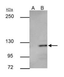
- Immunoprecipitation analysis of E-cadherin in MCF-7 whole cell extracts with E-cadherin polyclonal antibody (Product # PA5-85088) using 5 µg of sample, followed by anti-Rabbit IgG secondary antibody.

Supportive validation
- Submitted by
- Invitrogen Antibodies (provider)
- Main image
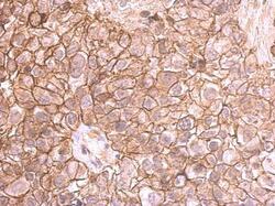
- Experimental details
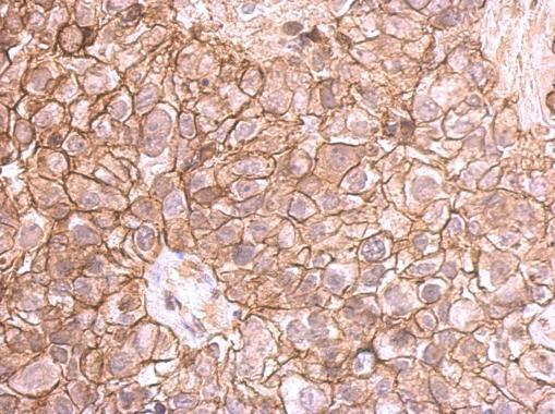
- Immunohistochemistry analysis of E-cadherin in paraffin-embedded breast cancer cells using E-cadherin polyclonal antibody (Product # PA5-85088) at a dilution of 1:500.
- Submitted by
- Invitrogen Antibodies (provider)
- Main image
- Experimental details
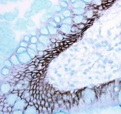
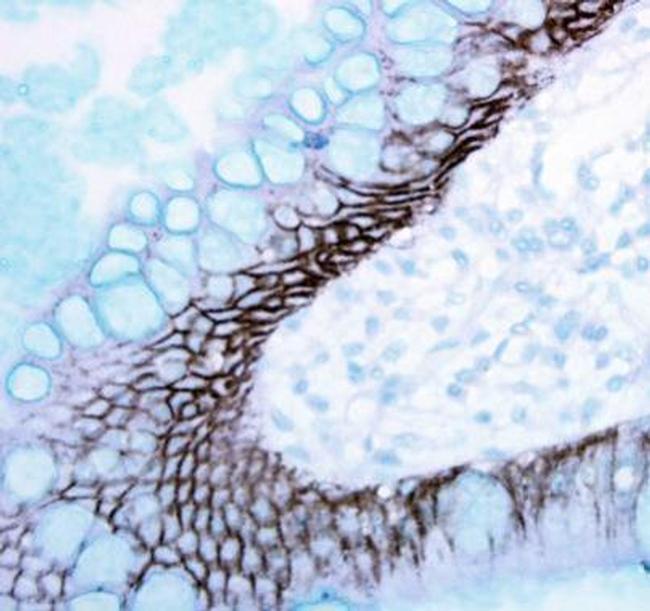
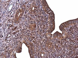
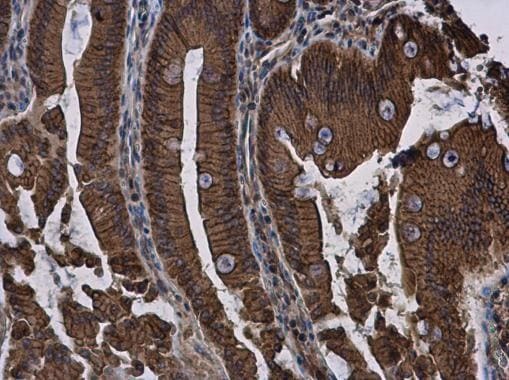
- Immunohistochemistry analysis of E-cadherin in paraffin-embedded human ulcerative colitis tissue using E-cadherin polyclonal antibody (Product # PA5-85088).
- Submitted by
- Invitrogen Antibodies (provider)
- Main image
- Experimental details
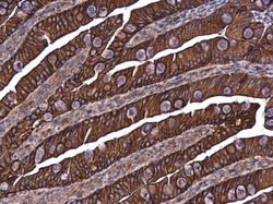
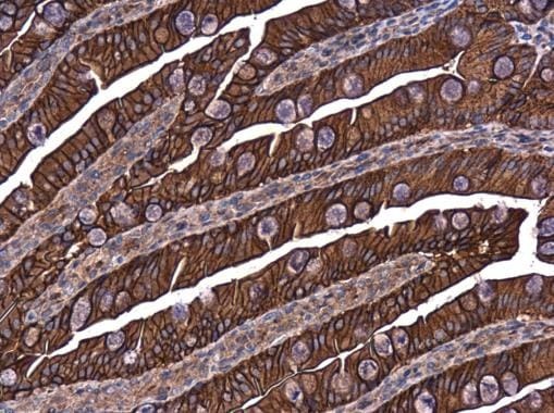
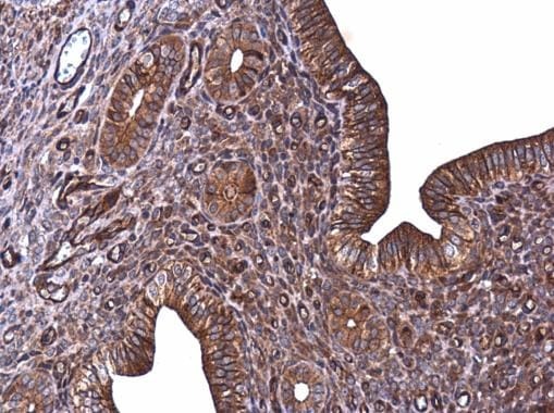
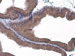
- Immunohistochemistry analysis of E-cadherin in paraffin-embedded rat intestine using E-cadherin polyclonal antibody (Product # PA5-85088) at a dilution of 1:500.
- Submitted by
- Invitrogen Antibodies (provider)
- Main image
- Experimental details
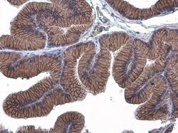
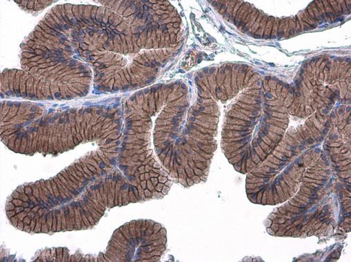
- Immunohistochemistry analysis of E-cadherin in paraffin-embedded rat prostate using E-cadherin polyclonal antibody (Product # PA5-85088) at a dilution of 1:500.
- Submitted by
- Invitrogen Antibodies (provider)
- Main image
- Experimental details
- Immunohistochemistry analysis of E-cadherin in paraffin-embedded mouse cervix using E-cadherin polyclonal antibody (Product # PA5-85088) at a dilution of 1:500.
- Submitted by
- Invitrogen Antibodies (provider)
- Main image
- Experimental details
- Immunohistochemistry analysis of E-cadherin in paraffin-embedded mouse pancreas using E-cadherin polyclonal antibody (Product # PA5-85088) at a dilution of 1:400.
- Submitted by
- Invitrogen Antibodies (provider)
- Main image

- Experimental details
- Immunohistochemistry analysis of E-cadherin in paraffin-embedded rat duodenum using E-cadherin polyclonal antibody (Product # PA5-85088) at a dilution of 1:500.
- Submitted by
- Invitrogen Antibodies (provider)
- Main image
- Experimental details
- Immunohistochemistry analysis of E-cadherin in paraffin-embedded rat prostate using E-cadherin polyclonal antibody (Product # PA5-85088) at a dilution of 1:500.

Supportive validation
- Submitted by
- Invitrogen Antibodies (provider)
- Main image
- Experimental details
- Immunoprecipitation analysis of E-cadherin in MCF-7 whole cell extract, A) control of preimmune rabbit IgG, B) immunoprecipitation of E-cadherin protein with E-cadherin polyclonal antibody (Product # PA5-85088) using A and B) 3 µg of sample at a dilution of 1:500. Sample was then incubated with anti-rabbit IgG secondary antibody. Prior to incubation with primary antibody, the sample was separated on 5% SDS-PAGE.

- Submitted by
- Invitrogen Antibodies (provider)
- Main image
- Experimental details
- Immunoprecipitation analysis of E-cadherin in MCF-7 whole cell extracts with E-cadherin polyclonal antibody (Product # PA5-85088) using 5 µg of sample, followed by anti-Rabbit IgG secondary antibody.
- Submitted by
- Invitrogen Antibodies (provider)
- Main image
- Experimental details
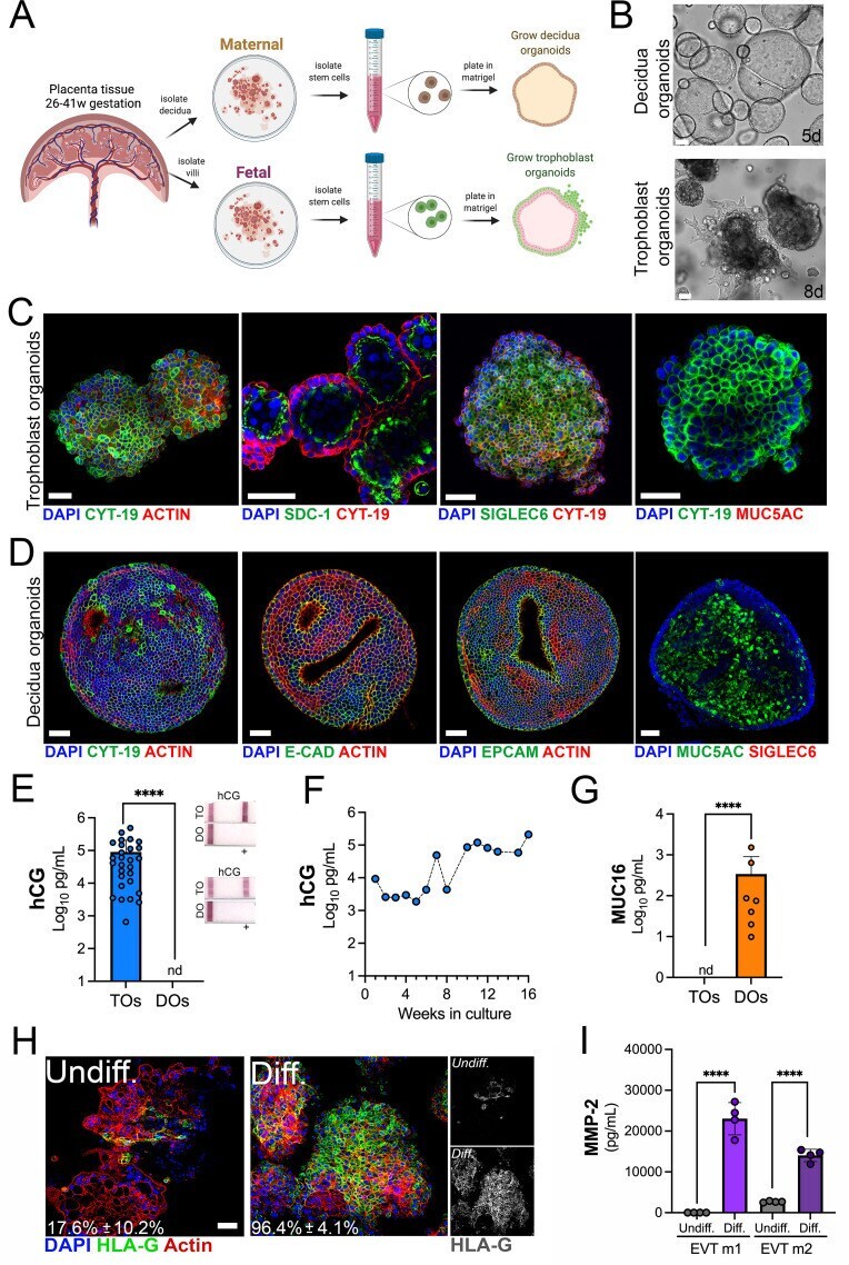
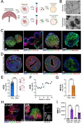
- Figure 1. Long-term three-dimensional organoids cultures can be established from human mid- and late-gestation placental tissue. ( A ) Schematic representation of the derivation of trophoblast organoids (TOs) and decidual organoids (DOs) from mid-to-late gestation placental tissue. ( B ) Representative bright-field images of TOs and DOs after passaging in complete growth medium TOM and ExM, respectively at 5 days (DO, top) or 8 days (TO, bottom) post-passaging. Scale bar, 50 um. ( C ) Confocal microscopy in TOs immunostained for (from left) cytokeratin-19, SDC-1, SIGLEC-6, cytokeratin-19 (in green) and actin, cytokeratin-19, or MUC5AC (in red). DAPI-stained nuclei are shown in blue. Individual channels are shown in Figure 1--figure supplement 1D . Scale bar, 50 um. ( D ) Confocal microscopy in DOs immunostained for (from left) cytokeratin-19, E-cadherin (E-cad), EpCAM, or MUC5AC (in green) and actin or SIGLEC6 (in red). DAPI-stained nuclei are shown in blue. Individual channels are shown in Figure 1--figure supplement 1D . Scale bar, 50 um. ( E ) Levels of hCG in conditioned medium (CM) isolated from TOs (blue) or DOs (orange) as determined by Luminex. At right, over the counter (OTC) pregnancy tests for hCG in two matched TO and DO lines. ( F ) Levels of hCG in CM isolated from TOs throughout a 16-week culture period as determined by Luminex. ( G ) Levels of secreted Mucin-16 (MUC16) in CM isolated from TOs (light blue) or DOs (orange) as determined by Luminex assays. ( H )